
Science is art: Imaging competition shows off the stunning side of microscopic research
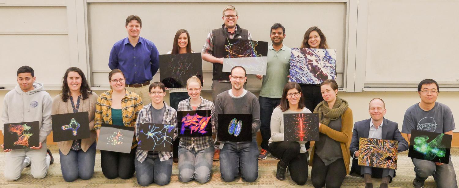
A world of hidden beauty exists all around us—if you just know where to look. Researchers from across CU Boulder illuminated the beauty of the microscopic realm for the CU Up Close Image Competition.
“The annual competition gives us a mechanism to demonstrate our imaging capabilities to a broad and diverse audience,” said Joe Dragavon, director of the BioFrontiers Advanced Light Microscopy Core. “It also encourages our researchers and investigators to take a step back from what may have become a daily activity, look at what they are actually doing, and realize that it is completely awesome and, in the case of optical imaging, stunningly beautiful.”
The contest, run by the BioFrontiers Institute, is open to researchers and investigators to help showcase the art that exists in science. Competitors capture still images and video of chemicals, organic matter, materials and more on BioFrontiers’ optical microscopes. Contest judges evaluate the submissions based on their aesthetic qualities and beauty. The judges select 12 images and three videos, and the public votes for the top three images and the top video.
"I think this contest is so important in so many ways,” said Leila Saleh, a graduate student in Chemical and Biological Engineering with the Bryant Research Group. “It's a misconception that science and art don't intersect. Scientists need creativity and originality to be successful just as much as artists do. I think we often lose sight of that when we get data-driven tunnel vision, so this contest serves a really great purpose in reminding us as scientists to step back and look at the bigger picture and the beauty of what we do. I think it's also a great way to get the general public involved in our work. Going to a talk can be intimidating, but a gallery of beautiful images draws you in."
The 2018 competition featured 75 images and videos submitted by nearly 40 researchers. The submissions came from undergraduates, graduate students, post-doctoral researchers and one faculty member. The competitors represented multiple departments and institutes.
From this pool, one first-place video and three images were chosen as the contest winners:
Video: 1st place: Quanbin Xu, Liu lab
Images:
- 1st place: Anastasia Karabina, Leinwand Lab
- 2nd place: Engineers and Scientists from Double Helix Optics, LLC
- 3rd place: Leila Saleh, Bryant Research Group
"Our light engineering technology is much like pointillism painting—we image each individual molecule in the sample and integrate those molecular-level images into the full picture you see in our photo,” said Warren Colomb of the DoubleHelix Optics team. “While this image is beautiful art, its real power lies in the scientific discovery enabled through its high-precision 3D dataset."
In addition to the top three images, nine more were selected for inclusion in a calendar showing off the top images from the contest. These 12 images are now on the walls of the third floor of the Jennie Smoly Caruthers Biotechnology Building on CU Boulder’s East Campus.
Dragavon said he hopes that these striking images will continue to help people to look at research in a new way.
“Much of what we do here at CU is absolutely amazing and incredible,” he said. “In the end, we are trying to remind our students that what they are doing is very impressive and that there is significant beauty in their science.”
Although past contests had a one-month entry window, the 2019 contest submission period is ongoing. Aspiring scientist-artists can submit for the contest right now.