Fall 2018 STEMinar Schedule
Current Academic Year (Fall 2018-Spring 2019)
Fall 2018
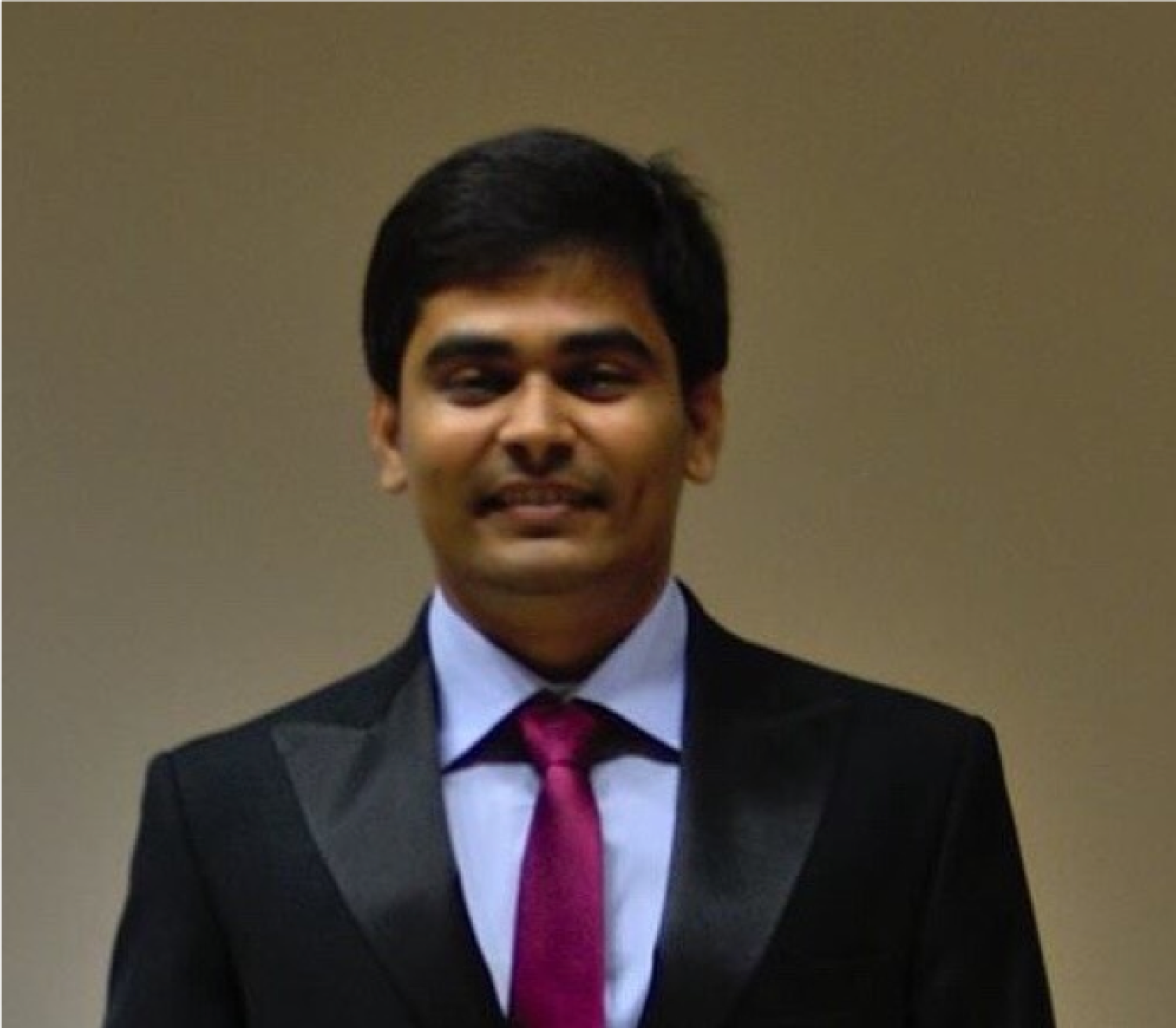
Speaker: Bhupendra Mishra
Title: "How Does a Blackhole Eat?"
Department: Astrophysical
Date: September 13th, 2018
Location: Duane Physics Reading Room (11th Floor, tower)
Time: 5:00 pm MST
The black holes are one of the most promising central objects to learn about the extreme physics involved in nature. They range from stellar mass to supermassive black holes. These monsters of the universe swallow everything coming close to it. The stars fall into black holes through giant disks around them. This is because the stars orbit around these black holes. The day to day life physics does not hold in the vicinity of these central objects and hence one needs to apply general relativistic theory proposed by A. Einstein. I shall talk about process through which the plasma from donor stars accrete into black holes. I shall give an overview of how black holes are formed and how do they shape the galaxies. The computer models will illustrate the challenging physics in these objects with the help of few simple movies I created.
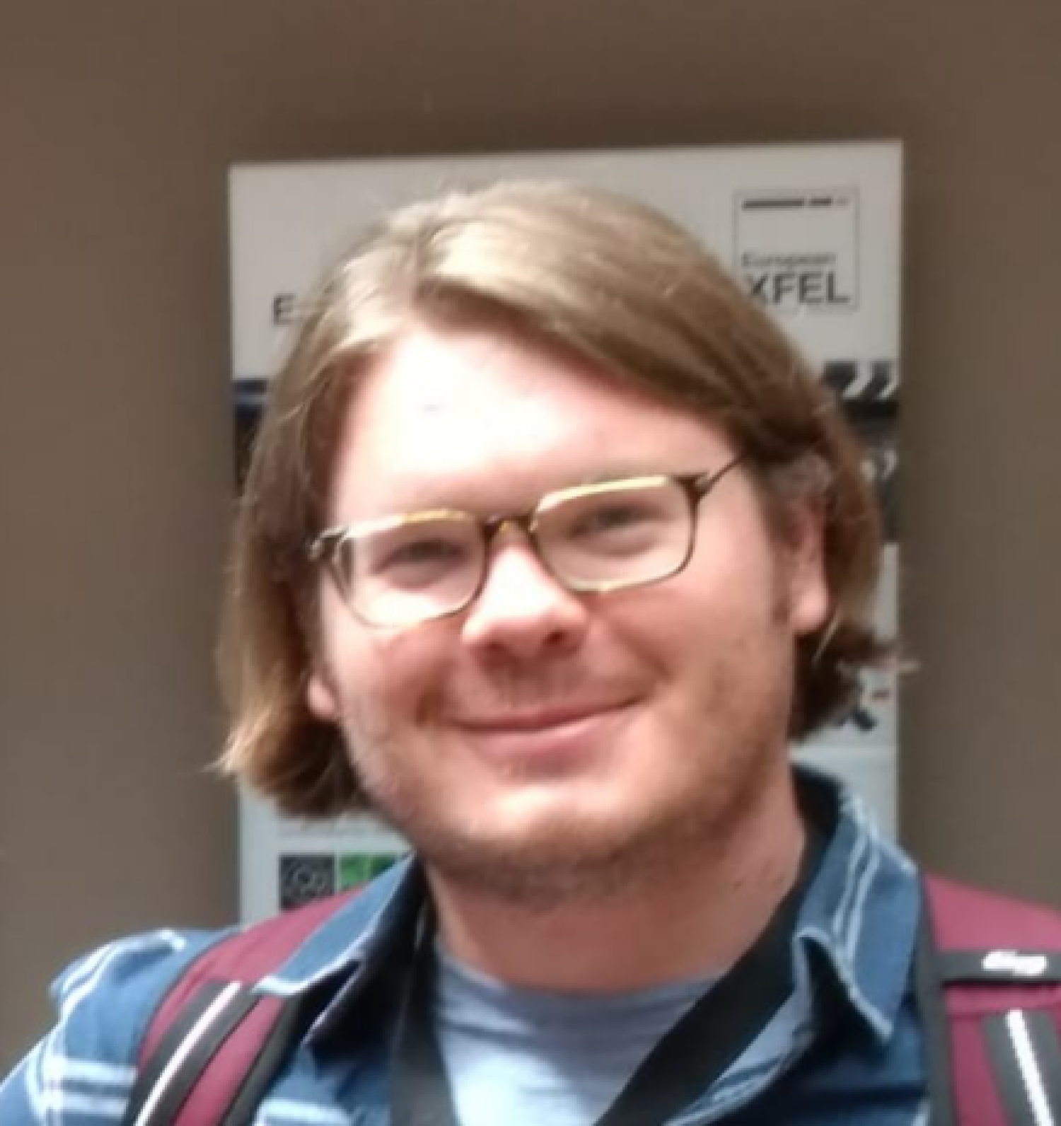 Speaker: Charles Bevis

Title: "What's going on at the bottom? Visualizing the nano-scale with lensless microscopy"
Department: Imaging Science (AMO Physics)
Date: September 27th, 2018
Location: Duane Physics Reading Room (11th Floor, tower)
Time: 5:00 pm MST
Have you ever wondered what's going on when things get very small? Well, when you're talking about the nano-scale that's not such an easy question to answer, and even harder to visualize. To resolve such small objects we need to use very short wavelength light, such as X-rays. Unfortunately, we can't make lenses for this kind of light. Which leads to the interesting problem: how do you form an image without a lens? The short answer is, of course, computers. The long answer requires a fascinating multidisciplinary approach that developed into a multi million-dollar industry and academic thrust. Developments in this field caused the rapid development of exotic new materials, machines, and methods to functionally solve a nonlinear problem that, in many cases, has no analytic solution. Come hear about the exciting field of computational imaging science and learn about the fundamentals of frequency space, Fourier transforms, and lensless nano-imaging!
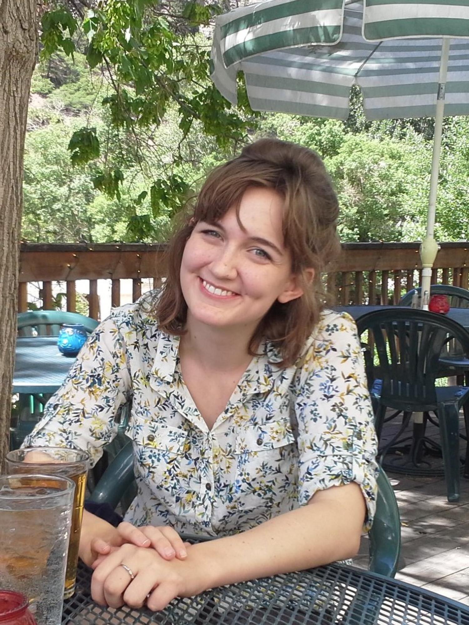
Speaker: Graycen Wheeler
Title: "Understanding cell signaling to fight cancer drug resistance"
Department: Biochemistry
Date: October 11th, 2018
Location: Duane Physics Reading Room (11th Floor, tower)
Time: 5:00 pm MST
Although cancer researchers have made huge strides in oncology therapies in the last several decades, many tumors are able to develop resistance to drugs and quickly come back after treatment. Understanding the processes that allow cancer cells to become resistant to commonly-used drug treatments will allow researchers to develop more effective cancer therapies and create better outcomes for patients. Cancer cells can develop drug resistance in many ways. For example, many cancer drugs work by damaging the DNA or the structural elements of a cancer cell so badly that the cell undergoes programmed cell death. Cancer cells can overcome this treatment by deactivating the cellular processes that sense this damage and signal for cell death. Cancer cells can also evade drug effects by making cellular machinery that pumps drugs out of the cell or by expressing proteins that cause the cell to grow and divide rapidly enough to counteract any drug-induced death.
My work focuses on one protein, CLPTM1L. Cancer cells make more of this protein than normal cells, and cancer cells that are resistant to several types of drugs make even more CLPTM1L than their drug-sensitive counterparts. If we prevent cancer cells from expressing this protein, we can kill them more quickly using significantly lower drug doses. I measured how much of this protein cells made after treatment with 96 FDA-approved cancer drugs and found that cells respond to treatment with many of these commonly-used therapies by making more of this protein. Since cancer cells express more CLPTM1L after drug treatment, and since this protein helps cancer cells survive drug treatment, CLPTM1L may be preventing many cancer drugs from reaching their maximum efficacy. I’m working to understand how cells know to upregulate CLPTM1L after drug treatment and how CLPTM1L helps cells survive that drug treatment. This research could allow the drugs we already use to treat cancer patients to be more effective at lower doses.
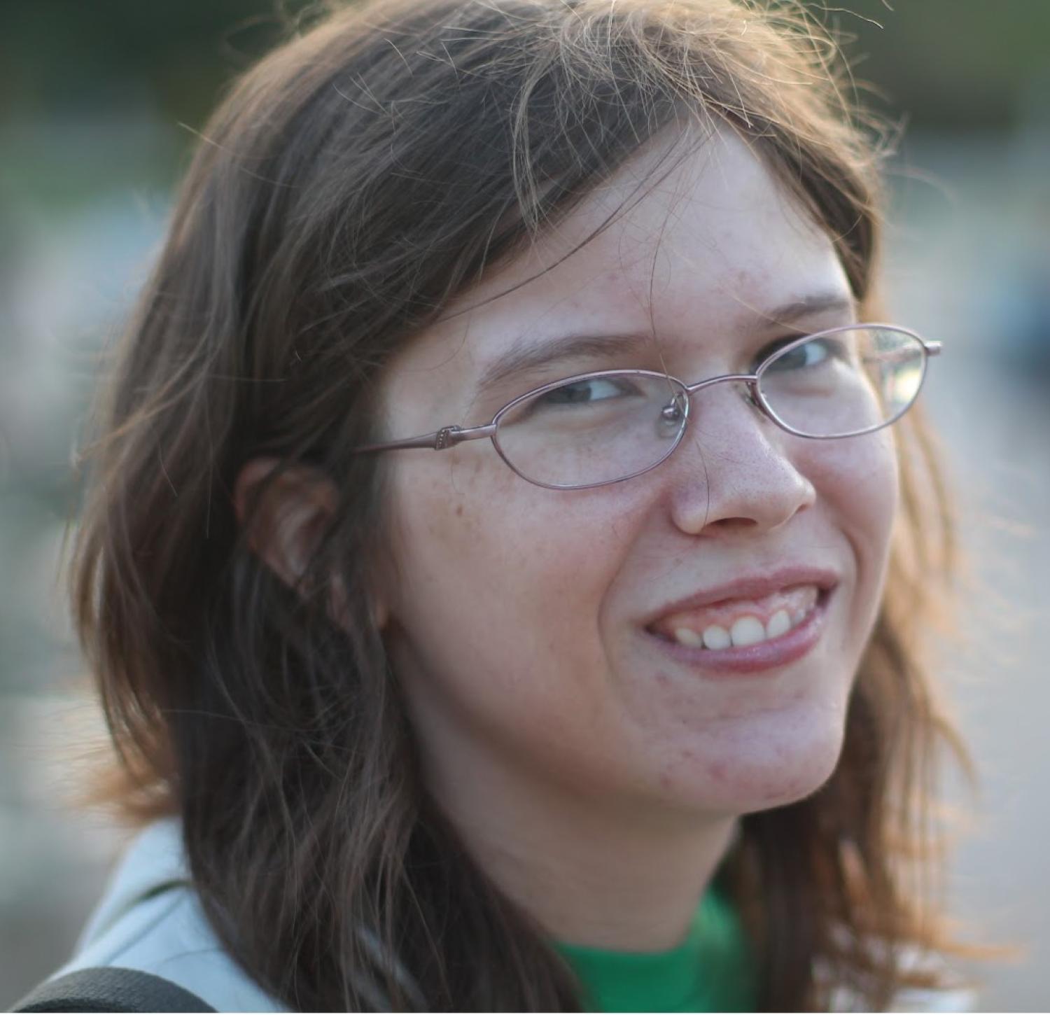
Speaker: Laura McGuire
Title: "Making a Universal Filter"
Department: Biophysics
Date: October 25th, 2018
Location: Duane Physics Reading Room (11th Floor, tower)
Time: 5:00 pm MST
Filters of many types are common in our everyday lives. The most common filters, like sieves, separate objects based on features such as size or electric charge, making it difficult to easily separate one species of molecule from a complex mixture. There is a need for a highly-specific, tunable filter that could easily select from among many different molecules. I work to create such a filter, taking advantage of the manner in which cells move molecules in and out of their nucleus. To enter the nucleus, each molecule must pass through a nuclear pore, a channel filled with long, flexible proteins that fill the channel like molecular spaghetti. The nuclear pore is highly selective, only allowing a few specific molecules to pass through, but the mechanism of selectivity is unknown. I investigate the filter through mathematical modeling and through experiment. My results suggest that the flexible, “wiggly” nature of the spaghetti-like proteins that fill the pore is crucial to their ability to accept or reject specific molecules. In order to test this prediction, I create hydrogel-based materials that mimic the environment of the nuclear pore, then test their selectivity. My ultimate goal is to understand the nuclear pore well enough to scale it up from a nanoscale channel to a macroscopic bulk material. Applications for this unique filter could then range from tissue engineering to smart drug delivery.
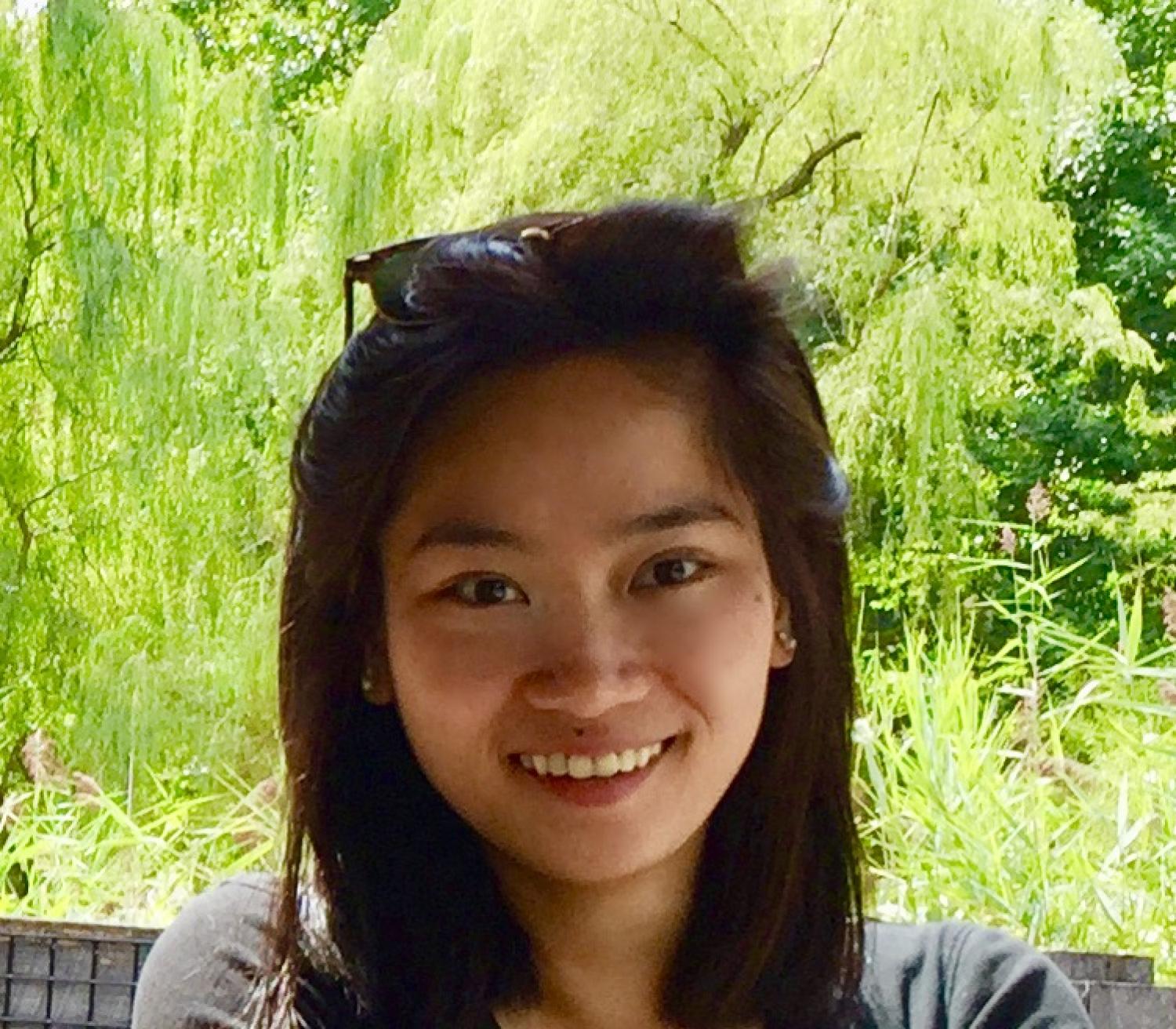 Speaker: Quynh Nguyen

Title: "Ultrafast Strong-Field Photoemission Dynamics of Ligand-free Nanoparticles in the Gas Phase with Visible to Mid-Infrared Laser Fields"
Department: Chemistry (Chemical Physics)
Date: November 8th, 2018
Location: Duane Physics Reading Room (11th Floor, tower)
Time: 5:00 pm MST
Over the past two decades, nanomaterials have grown from fundamental science endeavors to being present in our daily lives, owing to their unique nano-scale emergent phenomena. Nanomaterials have been used in a wide range of applications, including biofuels, biomedical technology, energy storage, as well as aerospace. In particular, the photophysical properties of nanomaterials have garnered great interest due to their tunability in composition, size, or geometry. However, these properties are not well understood due to the complex surrounding environments. Here, we overcome this limitation by combining a magnetron sputtering source with a velocity map imaging spectrometer, which allows us to study the photophysical properties of ligand-free nanoparticles, which are produced in the gas phase and not covered by layers of molecules. For the first time, we observe strong-field phenomena that occur at intensities orders of magnitude below those required to observe similar effects in atomic and molecular systems. Additionally, we uncover different ionization mechanisms that occur on picosecond (10-12 s) to femtosecond (10-15 s) timescales, which are dependent upon the nanoparticle composition. As a last thrust, we also present preliminary results on time-resolved photoelectron measurements of these nanoparticles, which opens the door to observing ultrafast nanoparticle photophysics on the attosecond (10-18 s) timescale. Taken together, the unique capabilities afforded by combining a magnetron sputtering source and a velocity map imaging spectrometer extends the methods of gas-phase atomic and molecular physics and attosecond science to the nanoscale materials. This will enable exciting applications such as ultrafast imaging of nano-scale potentials, state-resolved photoionization measurements, and surface plasmon dynamics and control.
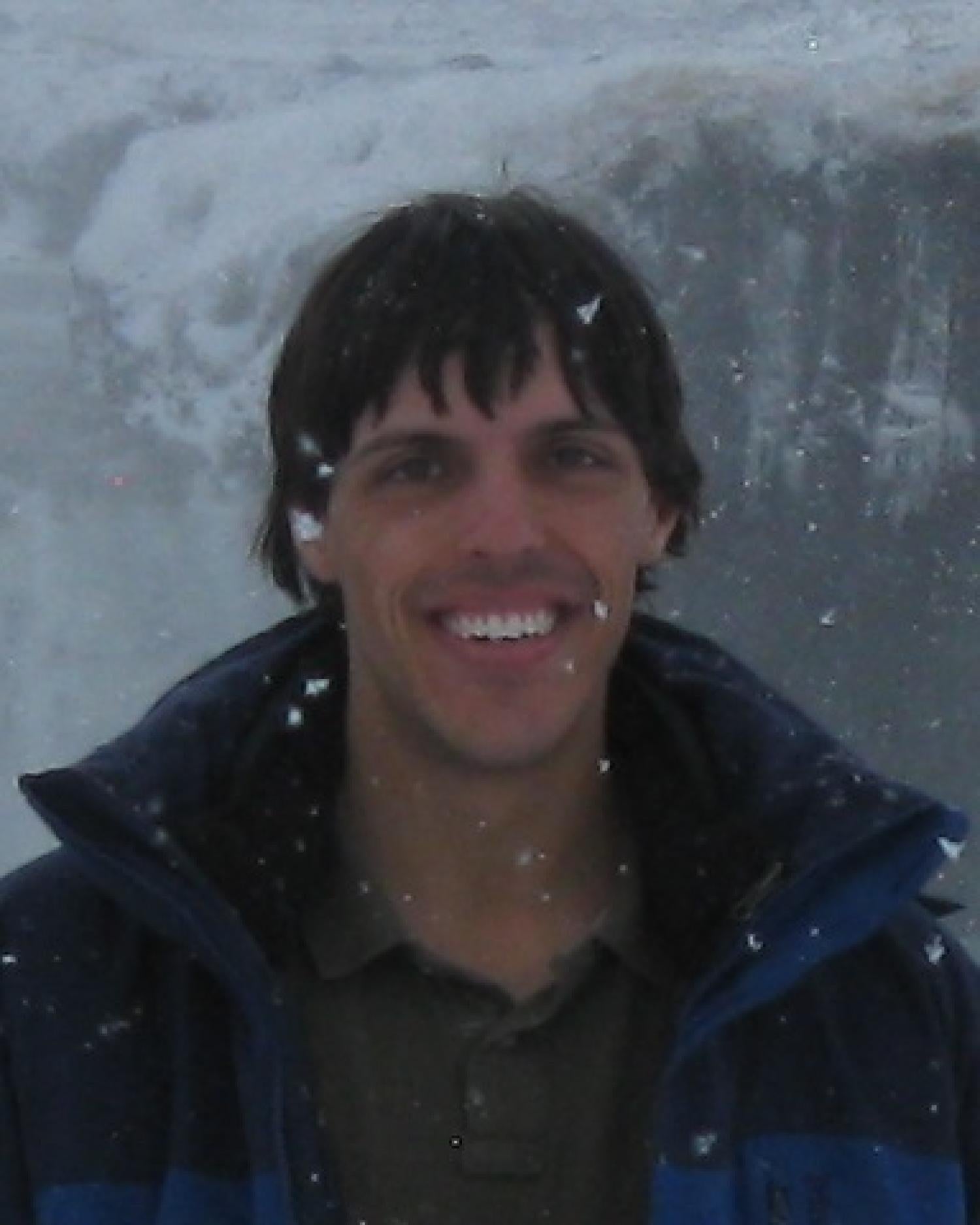
Speaker: Michael Tanksalvala
Title: "Coherent Diffractive Imaging: A Primer to Lensless Imaging"
Department: Electrical Engineering
Date: November 29th, 2018
Location: Duane Physics Reading Room (11th Floor, tower)
Time: 5:00 pm MST
Coherent diffractive imaging (CDI) comprises a set of imaging techniques that replace image-forming optics by a wide array of computer algorithms that retrieve an image from the scatter pattern generated by a coherent illumination beam.2, 3 CDI is particularly attractive for imaging with Extreme Ultraviolet (EUV) Light — EUV optics are expensive and difficult to fabricate, which precluded high-resolution, diffraction-limited EUV imaging to date. This talk will give an introduction to coherent diffractive imaging, as well as an overview of many applications in which it has been useful so far. I’ll talk about several of the major phase retrieval methods, including ptychography - a powerful implementation of CDI in which a beam is scanned region-by-region over a sample, and the collected diffraction patterns are combined to form a full-field, high-resolution, high-fidelity, complex image of the sample, with robustness ensured by the overlap between scan regions.1-4

