
FEATURED TECHNOLOGY
Advanced Infrastructure for Stem Cell Research
The Stem Cell Core Facility provides state-of-the-art infrastructure to support basic, translational, and regenerative medicine research. Our platform is designed to enable high-quality hiPSC maintenance, gene editing, molecular validation, and advanced imaging within a rigorously controlled environment.
🔬 Advanced Imaging & Cellular Characterization
High-resolution imaging platforms support morphological analysis, pluripotency validation, and immunofluorescence studies.
📌 Impact: Imaging platforms that directly support high-impact publications.
🔹 Olympus IX83 Microscope
High-resolution, fully automated observation of live and fixed samples across multiple dimensions (X, Y, Z, and time), supporting stem cell, organoids, embryos, and tissue research.
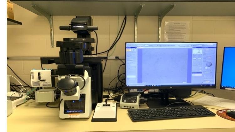

Key Features
- Z-Stack Imaging: Acquire images at multiple focal planes for 3D reconstruction
- Automatic Focusing: Multiple objective lenses for precise, reproducible imaging
- Time-Lapse Acquisition: Continuous monitoring of dynamic processes in live cultures, organoids, embryos, and organotypic slices
- Multimodal Imaging: Bright field, phase contrast, and fluorescence (DAPI, FITC, TRITC)
- Fast Image Acquisition & Overlay: View multiple fluorescence channels simultaneously
- Multipoint Scanning: Automated imaging at multiple locations for quantitative, reproducible datasets
🔹 EVOS M7000 Microscope
Fully automated fluorescence and transmitted-light imaging for live cells, organoids, embryos, tissue slices, and more. Designed for high-content stem cell research, these platforms combine speed, precision, and ease of use.
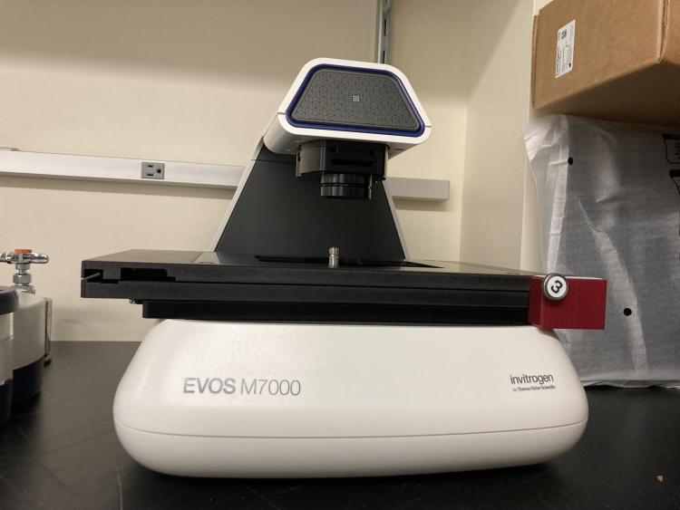

Key Features
- Automated Multichannel Fluorescence Imaging: Rapid, reproducible immunofluorescence data acquisition across multiple channels
- High-Quality Optics: 5-position objective turret with 4-color LED fluorescence and transmitted light channels
- Dual Camera System: 3.2 MP CMOS color and black/white cameras for bright-field and fluorescence imaging
- Automated Imaging: Fully automated X/Y scanning stage, autofocus, exposure control, and objective/light cube switching
- High-Speed Acquisition: Z-stack, tile-stitching, 2D/3D deconvolution, and time-lapse imaging for long-term experiments
- Environmental Control: Temperature, gas, and humidity regulation for live cell imaging
- Advanced Analysis: Optional Celleste™ software for cell segmentation, quantification, and visualization
- Flexible Workflow Programming: Well-plate scanning, multipoint time-lapse experiments, and montage/area scans in Z-stack or time-lapse modes
- Smart Illumination: LED technology ensures consistent fluorescence excitation with minimal phototoxicity
- User-Friendly: Intuitive interface for reliable operation across research groups
- https://www.thermofisher.com/us/en/home/life-science/cell-analysis/cellular-imaging/evos-cell-imaging-systems/models/evos-m7000.html
🔹 Olympus Provi-scanner
Digital whole-slide scanning for histological and immunostained samples.

Key Features & Benefits:
- Automated Slide & Culture Scanning: Rapid acquisition of multiple points in culture vessels or slides with minimal hands-on time.
- Non-Invasive Live Monitoring: Track cell health, confluency, and growth dynamics without staining or disrupting cultures.
- Remote Alerts: Receive notifications when cultures approach confluency and are ready for passage.
- High-Resolution Imaging: Bright-field, phase contrast, and fluorescence (DAPI, FITC, TRITC) for publication-quality data.
- Multichannel & Z-Stack Imaging: Capture multiple fluorescent markers, tile-stitch large areas, and generate 3D reconstructions.
- Data Management & Analysis: Easily store, reuse, transfer, and compare data across experiments and conditions; integrates with downstream analysis software.
- Versatile Sample Support: Histological sections, immunofluorescence slides, organoids, and tissue slices.
With fully automated imaging and live culture monitoring, the Olympus Provi-scanner enables quantitative, reproducible, and high-content data collection, empowering researchers to accelerate stem cell research with confidence.
🧬 Gene Editing & Genetic Engineering
Precision Genome Manipulation in hiPSCs, derived-Stem Cells and Primary Cell Cultures.
📌 Impact: These systems enable generation of isogenic lines, knockouts, knock-ins, and reporter cell lines.
🔹 Neon NxT Electroporation System
“Optimized for hiPSCs, primary cells, and other hard-to-transfect cell types"
- High Transfection Efficiency
- Customizable Protocols: Adjustable voltage, pulse width, and number of pulses for precise control
- Rapid & Reproducible: Consistent results across experiments and multiple users
- Versatile Applications: Supports CRISPR/Cas9 editing, plasmid delivery, RNA transfection, and protein introduction
- Minimal Cytotoxicity: Optimized settings reduce cell stress and maintain pluripotency
- Multi-User Ready: Reliable platform for academic core facilities and collaborative projects
- Seamless Workflow Integration: Compatible with downstream molecular assays, imaging, and functional studies
- Accelerates Research: Enables efficient genetic modification to support studies in differentiation, disease modeling, and regenerative medicine
🔹 Nucleofector 2b
Proven platform for CRISPR/Cas9 delivery and stable genome editing workflows.

- Efficient transfection of hard-to-transfect cell lines and primary cells.
- Easy adjustment and configuration of parameters.
🧪 Molecular Characterization & Validation
From DNA to Functional Assays
📌 Impact: These tools support pluripotency validation, differentiation markers, and genetic screening workflows.
🔹Real-Time PCR System
- QuantStudio 3 Real‑Time PCR System
- QuantStudio 5 Real‑Time PCR System

- Sensitive and Accurate Quantification: Detect and quantify DNA and RNA from stem cells, hiPSCs, and differentiated derivatives with high precision
- Fast & Reliable: Rapid cycling and optimized protocols for time-efficient gene expression and genomic assays
- Multiplexing Capability: Simultaneously measure multiple targets using fluorescent probes for complex stem cell pathways
- Wide Dynamic Range: Detect low-abundance transcripts, crucial for rare stem cell populations and differentiation studies
- Reproducible Results: Standardized workflows ensure consistency across experiments and users
- Easy Data Analysis: Integrated software for relative quantification, absolute quantification, and melt curve analysis
- Versatile Applications: Gene expression profiling, pluripotency marker validation, CRISPR genotyping, and stem cell differentiation studies
- Integration with Core Workflows: Compatible with upstream sample prep (RNA extraction, cDNA synthesis) and downstream validation assays
🔹Standard Thermocyclers
- ProFlex PCR System
- Prime PCR System


- Flexible Thermal Cycling: Multiple blocks and customizable profiles allow simultaneous amplification of different targets, ideal for multi-sample stem cell experiments
- High Precision & Accuracy: Uniform temperature control ensures reproducible amplification for DNA, cDNA, and genomic assays
- Multiplex Capability: Compatible with multiplex PCR assays for efficient detection of multiple gene targets in stem cells
- Rapid Cycling Options: Optimized protocols reduce experiment time without compromising data quality
- User-Friendly Interface: Intuitive software simplifies programming, monitoring, and data management
- Versatile Applications: Genotyping, pluripotency marker validation, CRISPR validation, and differentiation studies in hiPSCs and other stem cell lines
- Integration with Core Workflows: Works seamlessly with RNA/DNA prep, real-time PCR validation, and downstream molecular assays
- Reproducible Results Across Users: Ideal for multi-user core facilities requiring consistent and reliable data
🔹PCR Hood for DNA/RNA workflows

- Preparation of DNA and RNA samples for PCR, qPCR, and RT-PCR
- Setting up gene expression assays and genotyping workflows
- Minimizing contamination risk during sensitive nucleic acid handling
- Sample preparation for CRISPR validation and molecular cloning
Critical for maintaining data integrity in gene editing, pluripotency validation, and differentiation assays
🔹 Lucetta 2 Luminometer
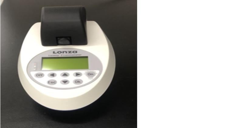
- Cell proliferation Assay.
- Cytotoxicity Assay.
- Luciferase reporter gene assay.
Mycoplasma Detection Assay.
🧫 Controlled Stem Cell Culture Environment
Reliable & Sterile Culture Infrastructure.
📌 Impact: This infrastructure ensures optimal sterility, cell viability, and reproducibility.
- Class II Biosafety Cabinets (5)
- HERAcell Vios 160i Incubathors
- Ultra-Low Temperature (ULT, -80*C) Freezer
- Microcentrifuges
- Water & Bead Baths
- Electronic Multichannel Pipettes
🔹 Ultra‑Low Temperature (ULT) Freezer

🔹Microcentrifuges

- Separation of cells, supernatants, and subcellular components
- Preparation of samples for PCR, RNA/DNA extraction, and enzymatic assays
- Pellet formation for cell washing, media changes, and differentiation protocols
Processing of small volumes for downstream analytical assays.
🔹Water bath & Beads bath
Water Bath

- Rapid thawing of cryopreserved cells and cryovials
- Warming media, reagents, and supplements
- Enzymatic incubations and enzyme activation steps
Temperature-sensitive molecular reaction
Bead Bath (Dry Bath)

- Safe, water-free alternative for thawing cryovials
- Warming media and reagents without contamination risk
- Incubation of samples in tubes
- Supporting standardized molecular and enzymatic workflows.
🔹 Electronic Multichannel Pipettes
Precision, Automation, and High-Throughput Performances.
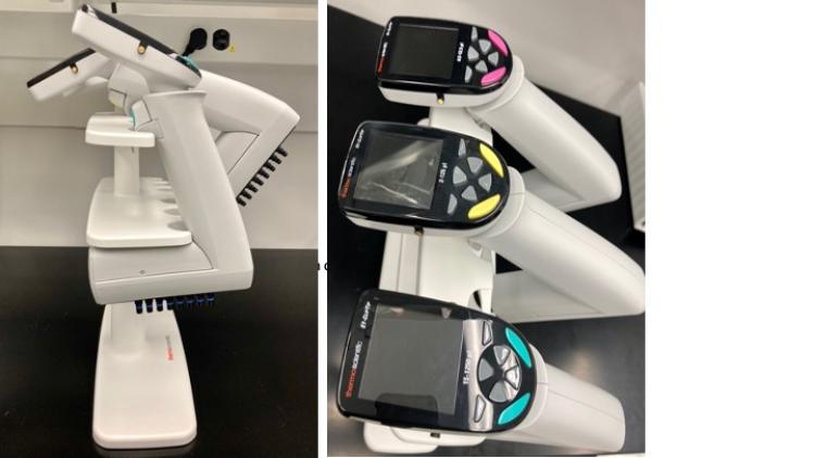
- High-throughput stem cell assays
- Standardized differentiation workflows
- CRISPR screening plate preparation
- Molecular assay reproducibility
🧬 Specialized hiPSC or derived-Stem Cells & WorkflowSupport
"Dedicated infrastructure to ensure optimal viability, morphology control, and reproducibility in stem cells workflows"
🔹 Sterile Dissection Hood + Olympus CKX53
For Colony picking/cleaning, stem cells picking/cleaning & morphology monitoring

- 4x, 10x, 20x, and 40x magnification.
- Bright field,
- Phase contrast,
- Fluorescence (DAPI, FITC, TRITC).
- Quickly acquisition and overlapping of images to view samples with multiple fluorescent lenses.
🔹 Automated Cell Counters:
- Countess II FL
- Countess 3

- Highly accurate and precise brightfield and fluorescence cell counting.
- Clump cell count.
- Automatic lighting and focus adjustment for optimal contrast and resolution.
- Automatic saving of cell counts and data.
- The software includes convenient built-in pre-dilution and cell-splitting calculators, especially for stem cell culture work.
Time to count two samples (sides A and B) ~1 minute
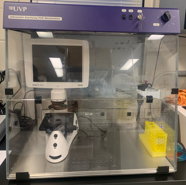
🔹 Sterilization Hood (UVP) + "Cell Culture Marker"

🔹 Automated Cell Thawing System (ThawSTAR CFT2)

➡ Standardized defrosting
➡ Reduces variability. Reproducible thawing profiles using a standardized thawing method.
➡ Ideal for cell banks
➡ Eliminate the risk of contamination https://www.youtube.com/watch?v=r2mOaIrt9Cw
🧫 Controlled Stem Cell Culture Environment
Equipment that supports cell maintenance, functional assays, and experimental stability.
🔹 ThermoMixer C (Interchangeable Thermoblock – Eppendorf)
** SmartBlocks for 15 ml tubes and plates

➡ Sample preparation
➡ Short incubations
➡ Enzyme assays
🔹 MicroPlate Centrifuge

➡ Rapid plate centrifugation
➡ ELISA, plate assays, PCR plates
🔹 CO2 Orbital Shaker Incubator
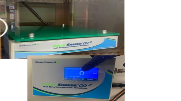
➡ Suspension cultures
➡ Organoids
➡ Differentiation under shaking.
🔹 Microsample Incubator

➡ Controlled short incubations
➡ Rapid assays
🔹 3D Orbital Mixer

➡ Gentle sample mixing
➡ Differentiation protocols
Imaging Support Platforms/ Training Platforms
🔹 Evos XL Core Microscopes
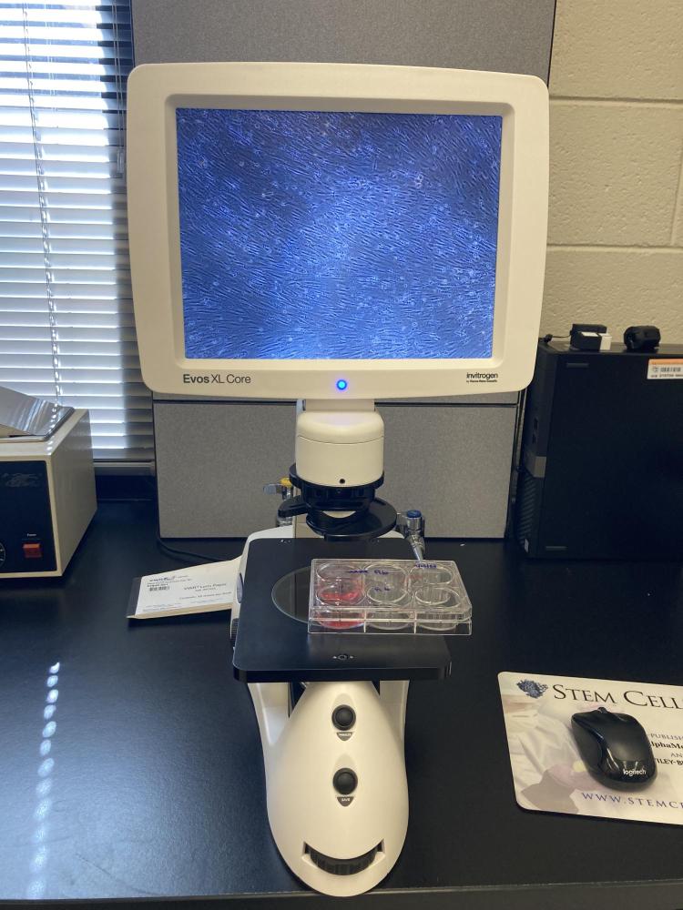
➡ Routine Microscopy. Objectives at 4x, 10x, 20x, and 40x.
➡ Daily Morphological Assessment
➡Easy image acquisition and storage during cell culture work.
➡ User Training
Biosafety cabinets with or without lab supplies and consumables.
For questions about lab equipment, please contact the Center!
__________________________________________________________________________________________
Videos:
Olympus IX83 Microscope
- IX83: Trans-mesenteric Migration of Enteric Neural Crest Derived Cells (ENCCs) https://www.olympus-lifescience.com/en/video/ix83_trans_mesenteric/
- IX83: Brain Slice Culture Glia Cell https://www.olympus-lifescience.com/en/video/ix83_brain_slice/
- IX83: Real-time Imaging for Ring1BYFP/YFP ME https://www.olympus-lifescience.com/en/video/ix83_ring1_byfp_yfp_me/
- IX83: Synthetic ERK Activity Propagation by a Light-Switchable System https://www.olympus-lifescience.com/en/video/ix83_synthetic_erk_activity_propagation/
- IX83: Light-induced Oscillatory/Sustained Expression of Ascl1 in Neural Progenitor Cellshttps://www.olympus-lifescience.com/en/video/ix83_neural_progenitor_cells/
- IX83 Inverted Microscope: Adding Oil to the Objective https://www.olympus-lifescience.com/en/video/ix83-inverted-microscope-adding-oil-to-the-objective/
- IX83 Inverted Microscope: Loading a Sample Slide on the Stagehttps://www.olympus-lifescience.com/en/video/ix83-inverted-microscope-loading-a-sample-slide-on-the-stage/
- IX83 Inverted Microscope: Focusing and Positioning the Stage Using the U-MCZ Remote Controllerhttps://www.olympus-lifescience.com/en/video/ix83-inverted-microscope-focusing-and-positioning-the-stage-using-the-u-mcz-remote-controller/