
Mircea Fotino, director of the Boulder Lab for high voltage electron microscopy, dies at 95.
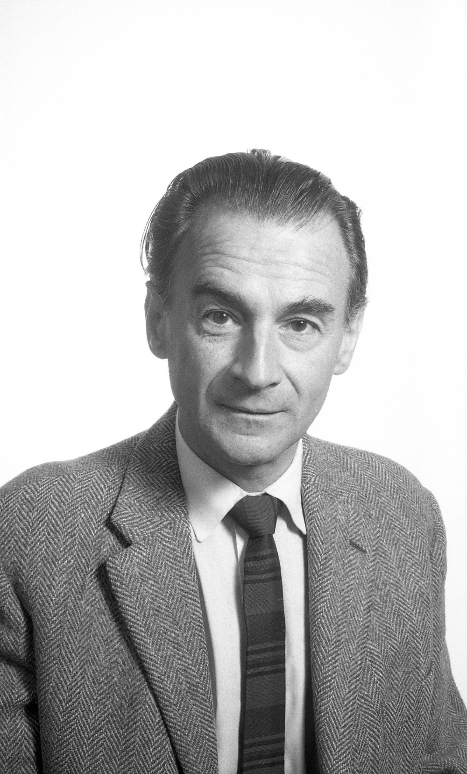
Mircea Fotino, a retired professor of Molecular, Cellular, and Developmental Biology (MCDB), passed away on January 23, 2023, dying peacefully at home in the company of his family. Mircea was born in Bucharest, Romania, in 1927, son of Scarlat Fotino and Marcella Teodorescu. He came to the USA for the latter part of his education, earning a Ph.D. in Physics at the University of California, Berkeley in 1958. He did postdoctoral work as Chargé de Research at the Centre National de la Research Science in Paris, France from 1959 – 1960, then came to Harvard University in Cambridge, MA, where he worked as a Research Fellow in Physics at the Cambridge Electron Accelerator from 1961 – 1968. Mircea married Ingrid Alina Popa in 1969, and they raised two daughters: Alina Domnica and Adriana Georgia.
Keith Porter, Chair of Biology at Harvard, convinced Mircea to accompany him to Boulder and help set up a facility for the study of biological samples by high voltage electron microscopy (HVEM), which Porter wanted to establish in the new biology department that was forming at CU Boulder. Mircea was a research fellow from 1968 – 1970, during which he explored the availability and use of such microscopes. In 1971, he was appointed Assistant Professor of MCDB and thereafter devoted his time to testing commercially available HVEMs. He traveled to England, France, Germany, Korea, and Japan where such microscopes were either available or under consideration. From these experiences, he learned the strengths and weaknesses of each microscope design. He also learned the many complexities of installing and running such a microscope. These were significant issues, because the electron accelerators that could bring a conventional electron beam (100 KV) up to the million volts needed for high voltage work required considerable space and mass, as well as high vacuums and large, effective insulators. Mircea selected the 1,000 KV microscope made by the Japanese Electron Optical Laboratories (JEOL) and returned to Boulder to supervise the construction of an appropriate home for the instrument.
Thanks to Mircea’s knowledge and care, the facilities necessary for the three story-tall microscope were built right into underground portions of the building now known as Porter Biosciences. His designs were so effective that this microscope, which weighed several tons, was efficiently installed, and soon thereafter was delivering images with resolutions measured in tenths of nanometers. Mircea served as director of the Boulder Lab for HVEM for over 30 years. He organized numerous workshops on the use of the instrument and supervised the work of visitors to this National Research Resource. Under his aegis, hundreds of thousands of micrographs were taken by scientists who wanted to learn what HVEM could do for the understanding of their scientific problem. Mircea built useful devices for aligning pairs of tilted views, so biologists could see their samples in stereo, and he worked with visiting biophysicists, like Professor Lee Peachy from the University of Pennsylvania, to help quantify their 3D images. Mircea also developed a course for MCDB and the Department of Physics on electron microscopy, the physics of electron microscopes and emerging subfields of biophysics.
Mircea’s personal research was a pioneering effort to use HVEM to visualize biological samples that were frozen, rather than fixed with chemicals. He believed, as did many other structural biologists, that rapid freezing was the most reliable method for accurate preservation of biological materials, at the same time making specimens able to withstand the high vacuums necessary for electron microscopy. Mircea hoped that the high energy of the electrons in an HVEM would penetrate the thin margins of whole cells, allowing visualization of structural details in cellular samples made using the best possible mode of preparation, thereby achieving electron optical resolution of well-preserved cellular details. Dr. Thomas Giddings worked with Mircea on this project for several years, and together they were able to get frozen cells into the HVEM, using a special low-temperature stage that Mircea had built. Initially, they were unable to see useful structural details in these thick and complex frozen-hydrated samples. They found, however, that if the samples were left in the microscope at temperatures above that of liquid nitrogen, much of the cellular water would sublime away, allowing examination of cells that had been freeze-dried at high vacuum and never exposed to air. Because of the complexity of this operation and the expense of the HVEM, this method was never widely used, but papers describing the work were published.
Selected papers:
Fotino M, Giddings TH. Ultrastructural visualization of unfixed and unstained whole mounts by high-voltage electron microscopy at low temperatures. J Ultrastruct Res. 1985 May;91(2):112-26. doi: 10.1016/0889-1605(85)90063-1. PMID: 4087332.
Fotino M. Electronic image formation in high-voltage TEM by sequential pixel acquisition. J Microsc. 1986 Feb;141(Pt 2):223-42. doi:10.1111/j.1365-2818.1986.tb02718.x. PMID: 3959073.
Fotino M. Visualization of biological specimens by cryoHVEM. J Microsc. 1986 Sep;143(Pt 3):283-98. doi: 10.1111/j.1365-2818.1986.tb02785.x. PMID: 3097320.
Fotino M. Micromorphology by cryo-HVEM. Prog Clin Biol Res. 1989: 295:581-7. PMID: 2748676.
Richard McIntosh, January 29, 2023