Imaging
Microbubble Ultrasound Contrast Agents
Microbubbles are the ultimate mechanically responsive colloidal particles. The compressible gas core and resonant response of the microbubble at MHz frequencies makes it the ideal ultrasound contrast agent and drug delivery vehicle. No other particle comes close to the acoustic echo – a single microbubble can be detected in vivo with current clinical scanners. Each microbubble is only 1-10 μm diameter and stabilized by a lipid shell, so they are safe for intravenous injection. In fact, they have been FDA approved for echocardiography for more than a decade. Ultrasound can also be used to push microbubbles (radiation force) and destroy them in the beam focus for targeted delivery. Our goal is to engineer innovative microbubble suspensions for imaging, therapy and theranostics (therapy + diagnostics). Our approach is to study the intermolecular and surface forces and interfacial transport processes that control the structure, properties and performance of microbubble suspensions, and to exploit these for rational design.
Read more here: (2002), (2004), (2018), (2020)

Microbubble shells often exhibit beautiful microstructures
Nanodrop Phase-Change Agents
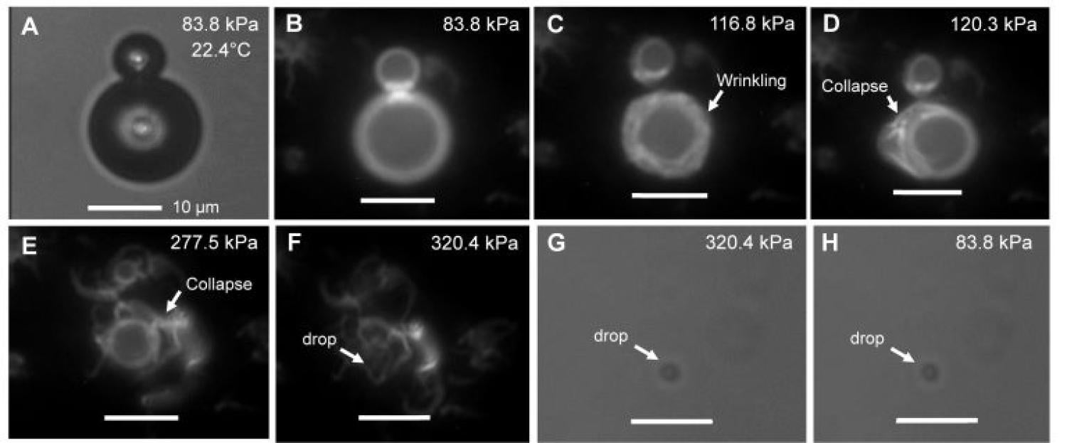
Nanodrops are condensed microbubbles – they offer the same acoustic effects as microbubbles, but in a much more compact form. A nanodrop can be formed by condensing the microbubble’s fluorocarbon vapor core into liquid. Once condensed, the nanodrops are remarkably metastable and remain in liquid form until activated by acoustic or optical energy. User-controlled activation stimulates conversion of the liquid core back into a vapor, transforming the nanodrop back into a microbubble. This is the hot new area in ultrasound contrast agent design! Again, our goal is to engineer nanodrops for innovative biomedical applications, and our approach is to focus on the relevant intermolecular and surface forces.
Read more here: (2014), (2015a), (2015b), (2016), (2017), (2018)

Nanodrops can be formed by pressure-induced microbubble condensation
Ultrasound Molecular Imaging
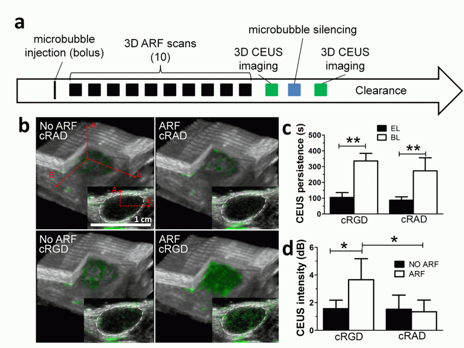
One of our goals is to engineer microbubbles and nanodrops to image the expression of key molecules associated with disease and the response to therapy.
Read more here: (2008), (2013), (2018a), (2018b), (2019), (2020)


