
The CU Museum is currently closed and will reopen in the fall of 2026!
Please email cumuseum@colorado.edu for more information.
The Double-Edged Sword of Digitization: New Technology, New Problems
Garret Jolma
Digitization is a crucially important part of every natural history museum’s work these days. Not only do museums house tens of thousands of specimens, but they are striving every day to turn those specimens into digital information that can be shared far and wide. Pictures of specimens can help scientists better identify species, and information from specimen labels can inform us how animals are reacting to climate change. Recently, the CU Museum of Natural history got a new tool to aid its digitization efforts: a camera and computer setup capable of creating three-dimensional models of insect specimens. This photography setup is the latest generation of imaging technology used in museum digitization. But in addition to providing scientists with valuable data about insects, this camera and the models it creates threaten to overload the Museum of Natural History’s digital storage systems.
Before the camera rolls, let’s set the stage: the entomology collection’s new camera is just part of a photography setup that also includes moving rails, spinning mounts, and synchronized flashes at every angle. The insect specimen, the star of our show, is placed on a circular mount. This mount can rotate, spinning the insect like a top (though not as quickly!). Surrounding the mount is a plastic light diffuser, meant to even out the flashes’ bursts of light across the insect. The entire mount and diffuser assembly is on a rail that can move closer or farther from the camera. And the camera is connected to a nearby computer so that pictures can be taken remotely. There are a lot of parts to this new setup, but it enables us in the entomology collection to take beautiful and useful pictures of our specimens quickly and efficiently.
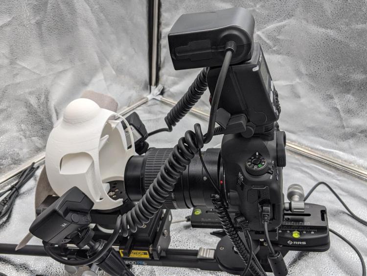
Figure 1: The Entomology collection’s new photography setup. The rotating mount is on the left, surrounded by the white plastic diffuser. Photo credit: Garret Jolma.
But why might a museum want to take pictures of its specimens? Why all the fuss for some dead bugs? The entomology collection’s new camera rig can take high-resolution magnified images of any insect specimen, and those sharp images have many uses. Entomologists and taxonomists can use those images to see if an insect belongs to a new species, based on the insect’s body features. Evolutionary biologists can use those images to see how insects’ body features change over time; they might even be able to tell if the imaged insects are adapting to climate change in some way. And, perhaps best of all, these scientists can do this valuable work without having to leave home. CU’s Museum of Natural History can send them the pictures and they can do this research from their offices.
This sharing of data is the idea behind digitization, the process of imaging museum specimens, their labels, or a collector’s field notes. Before an item is digitized, the only way to get data from it is to go to it and take measurements. Taking those measurements can be easier said than done, especially if you are a researcher who lives two states away from the item you want to see. But once that item is digitized, then you can take measurements of a bee’s legs or a butterfly’s wings from the digitized images. Done correctly, these images are at such high resolution that the results are no different—sometimes, even better—than taking measurements from the insect itself.
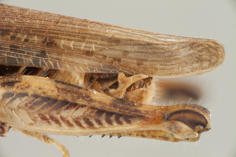
Figure 2: A close-up of a Little Spur-throated Grasshopper, Melanoplus infantilis. The forked protrusion on the grasshopper’s abdomen, just under the wing here, is diagnostic for this species. Specimen number UCMC 0051636. Photo credit: Garret Jolma.
Getting images of such high resolution isn’t as easy as aiming the camera and hitting “go” on the computer. The biggest problem we run into when photographing such small insects is how to keep the entire insect in focus at once. Let’s say we want to take a picture of a bee that is only one centimeter long. At those scales, the camera will be able to focus on the bee’s face, but not its abdomen; and if the camera focuses on the abdomen, the face will be out of focus. To solve this, we just take several pictures of the bee at different focal lengths: ten to thirty pictures, with different parts of the bee in focus in each picture. Then we feed the pictures into a computer program that can combine all the pictures into a single image where the entire bee will be in sharp focus. This is called focus-stacking. Not only are these focus-stacked images useful scientifically, but they are often beautiful to boot.
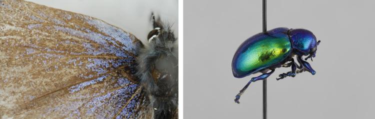
Figures 3 & 4: A close-up of a butterfly and a metallic beetle, both from the Entomology teaching collection. Photo credits: Garret Jolma.
What makes the entomology collection’s new photography setup special, however, is that it can turn these focus-stacked images into a 3D model of an insect specimen. With a pinch of math and some clever automated computer instructions, we can take focus-stacked images of our specimen from many different angles, slowly rotating the pinned insect to get images from all sides. Another computer program can turn these images, each taken from a slightly different angle, into a single 3D model of the insect specimen.
These 3D models are incredible. They allow researchers to get three-dimensional data from a museum specimen instead of just using a flat picture; they are the next best thing to actually handling the specimen. The only issue is that these 3D model files are quite large. Where a single picture from our camera is only a few megabytes in digital storage size, one 3D model can be ten times as large. It doesn’t take many 3D models to fill up a lot of storage space on a computer.
These large files are where CU’s Entomology collection is running into problems. Though our research tools are state of the art, our digital storage solutions are lagging behind. Currently, the Museum of Natural History has no integrated digital storage system. Each section—Anthropology, Botany, Zoology, and so on–is left to fend for themselves. Entomology’s current system of storing digital pictures is to load them onto external hard drives and then store the hard drives in the collection room or a side office. So far, this method of storing files has worked, if only barely.
However, there are many ways this method could go wrong, first and foremost being that CU Entomology only has single copies of its files. For now, we only have one copy for each image, so if something happens to an image, there is no backup. The best practice in this situation would be to have two copies: an archived copy and a regular use copy. While this solution would be ideal, it would also double our storage requirements—a hard-to-swallow solution for a museum section already struggling to find digital storage space.
The second issue is the longevity of the stored images. Currently, CU Entomology is storing its images as .jpegs, a common and easily-readable file type. But each time a .jpeg is opened, it degrades the image. The degradation isn’t noticeable on short time scales, but if we want these images to last as long as the pinned insects, we have much more work to do. To keep pictures for longer, they can be saved in an archival file format, like DNGs. DNGs do not lose data or quality when they are opened, ensuring that the files will last long into the future.
Finally come the physical concerns behind data storage. If we put our pictures on external hard drives, where should we put the hard drives? At the moment, they are in our collection manager’s office or on a bookshelf. These sound like safe places until you consider how old the Museum Collections building is. The Museum Collections Building (MCOL) was built about a hundred years ago to be the Geology Building, and was retro-fitted twenty years ago to house the museum’s collections. The Entomology section has dealt with blown-in windows and water leaks regularly in the past few years. Just this winter, a leak sprung in the ceiling of our collection manager’s office, threatening her bookshelf and everything on it. What good is an archival quality image if the hard drive it’s on gets destroyed by a leaky ceiling?
All the collections managers and curators at CU’s Museum of Natural History are in agreement about the solution to these problems: hiring a digital assets manager to construct and oversee a digital storage system for the museum. Then we would have a digital storage method commensurate with our high-caliber research. Other museums, such as the Field Museum in Chicago and the Denver Museum of Nature & Science, have an entire department dedicated to handling the data that is created in the process of doing research.
Despite the challenges, the Entomology collection on campus will continue its research, taking focus-stacked images of its specimens and crafting 3D models for researchers to share. Keep an eye on the Museum of Natural History’s online presence, and you may just see some of those images on your own screen.
Emerging Technologies Student Blogs
The Double-Edged Sword of Digitization: New Technology, New Problems
Garret Jolma