
Confocals
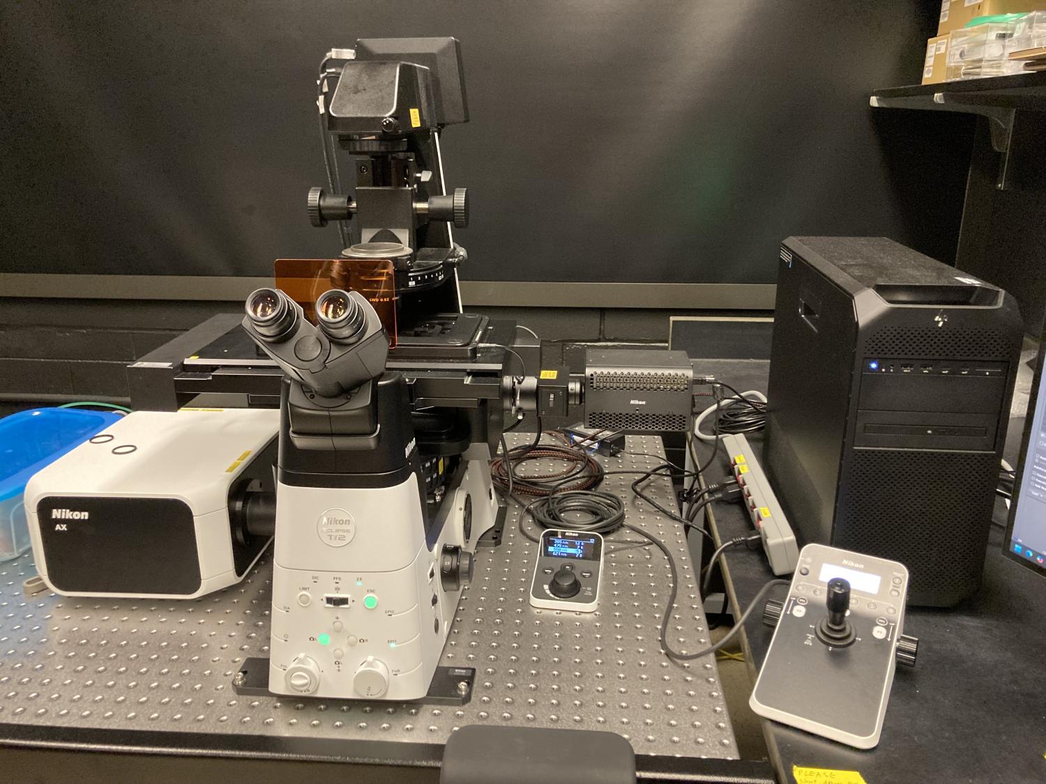
RED BARON - Nikon Ti2E AXR NSPARC laser scanning with resonance and super-resolution (Porter B049)
Lasers
Launch: LUD-S4
- Excitation at: 405, 488, 561, 640 (nm)
Emission filters & molecules imaged (All filters are Chroma)
Confocal scanning:
- Emission filter: 430-475 (nm); DAPI, Hoechst, DyLight-405, Alexa405, etc.
- Emission filter: 499-551 (nm); Alexa488, FITC, EGFP, Opal520, etc.
- Emission filter: 571-625 (nm); Alexa555, 568, 594, TxRed, mRFP, mCherry, TdTomato, Opal570, etc.
- Emission filter: 663-738 (nm); Alexa647, Cy5, Opal620, etc.
- DIC imaging (laser scanning confocal only, 10, 20, 40, 60x)
NSPARC scanning:
- Emission filter: 430-475 (nm); DAPI, Hoechst, DyLight-405, Alexa405, etc.
- Emission filter: 502-546 (nm); Alexa488, FITC, EGFP, Opal520, etc.
- Emission filter: 570-616 (nm); Alexa555, 568, 594, TxRed, mRFP, mCherry, TdTomato, Opal570, etc.
- Emission filter: 666-732 (nm); Alexa647, Cy5, Opal620, etc.
Light source, filters & viewing by eye
- Nikon D-LEDI; 385, 475, 550, 621 (nm)
- DAPI, GFP, RFP, Cy5 with quad-band emitter (Chroma)
- Brightfield
- DIC (10, 20, 40, 60x)
NSPARC super-resolution lens
- 60X PLAN APO TIRF oil NA 1.49 WD 0.12 (mm), DIC
Additional standard objectives
- 2x PLAN APO λ NA 0.10, WD 8500 (mm)
- 10X PLAN APO λ D NA 0.45 WD 4.0 (mm), DIC
- 20X CFI60 PLAN APO λ D NA 0.80 WD 0.80 (mm), DIC
- 40XC PLAN APO λ S SILICONE NA 1.25 WD 0.30 (mm), DIC, correction collar (23-37 degrees C)
- 60X PLAN APO IR SR water 1.27NA WD 0.18 (mm), DIC
Available upon request
- 60x 1.40NA Plan Apo OIL VC λ WD 0.13 (mm)
- 40x NA 0.95 WD 0.18 (mm)
- Inquire with James.Orth@Colorado.edu
Laser scanning
- DUX-VB, 4 PMT-GA GaAsP (photo-multiplier tubes)
Super-resolution
- Nikon Spatial Array Confocal (NSPARC) pinhole array sensor; 5x5 array of 0.2 AU pinholes
Sample Holders
- Slide, 35 (mm) dish, 50/60 (mm) dish
- Multi-well plate
Capabilities
- High frame-rate imaging using resonance scanner
- Super resolution to 100nm XY using NSPARC detector
- Queensgate (Prior QGNPS083-NI) Piezo Z-stage with 600 micron travel
- Adjustable GFP and RFP emission bandwidth (confocal only)
- TOKAI-HIT Cell Incubator 1 x 35 , 50 or 60(mm) dish, chamber side (coverslip bottom), multiwell plate
- Motorized X, Y
- Multi-point, time-lapse, optical sectioning, large-area montage with stitching
- Advanced analysis tools via separate HP Z4 workstation, including spectral deconvolution, and full analysis suite (inquire with Director)
- Nikon Perfect Focus System
- NIS Elements AR 6.02.03 with JOBS, general analysis
Email, James.Orth@Colorado.edu
Reserve the Ti2E AXR NSPARC LSC
For training email, James.Orth@Colorado.edu
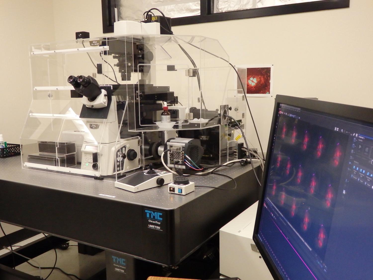
THOR - Nikon TiE A1 laser scanning with structured illumination and TIRF (Porter B047A)
Lasers
Launch: LUNV6, high-output
- Excitation at: 405, 445, 488, 514, 561, 647 (nm)
Emission filters & molecules imaged (All filters are Chroma)
- Emission filter: 425-475 (nm); DAPI, Hoechst, DyLight-405, Alexa405, etc.
- Emission filter: 467.5-502.5 (nm); CFP, cerulean, etc.
- Emission filter: 500-550 (nm); Alexa488, FITC, EGFP, Opal520, etc.
- Emission filter: 521.5-554.5 (nm); YFP, Venus, etc.
- Emission filter: 575-625 (nm); Alexa555, 568, 594, TxRed, mRFP, mCherry, TdTomato, Opal570, etc.
- Emission filter: 650-720 (nm); Alexa647, Cy5, Opal620, etc.
- DIC imaging possible (laser scanning confocal only, 20, 40, 60, 100x)
Light source, filters & viewing by eye
- Lumencor SOLA
- DAPI, GFP, RFP (Chroma)
- Brightfield
- DIC (20, 40, 60, 100x)
N-SIM super-resolution lens
- 100x 1.49NA OIL SR Apo TIRF
Additional standard objectives
- 4x 0.2NA Plan Apo λ WD 20 (mm)
- 10x 0.45NA Plan Apo λ WD 4 (mm)
- 20x 0.75NA Plan Apo VC λ WD 1 (mm)
- 40x 0.95NA Plan Apo λ WD 0.25-0.17 (mm)
- 60X 1.27NA Water SR Plan Apo WD 0.17 (mm)
Available upon request
- 60x 1.40NA Plan Apo OIL VC λ WD 0.13 (mm)
- Inquire with James.Orth@Colorado.edu
Laser scanning
- 2 x GaAsP high-sensitivity PMTs (CFP, GFP, YFP, RFP), photo-multiplier tubes
- 2 x multi-alkali PMTs (DAPI, far-red), photo-multiplier tubes
N-SIM & TIRF
- Andor iXON 897 Ultra EM-CCD (512x512)
Sample Holders
- Slide, 35 (mm) dish, 50/60 (mm) dish
- Multi-well plate
Capabilities
- 3D structured illumination, 'fast' 2D SIM
- TIRF
- TOKAI-HIT Cell Incubator 1 x 35 (mm) dish, chamber side (coverslip bottom)
- Motorized X, Y, Z and separate MCL Piezo Z with 200 micron range
- Multi-point, time-lapse, optical sectioning, large-area montage with stitching
- Advanced analysis tools via separate workstations, including spectral deconvolution, and full analysis suite (inquire with Director)
SIM imaging with 647nm laser:
- NIS Elements AR 5.04; 64 bit
Email James.Orth@Colorado.edu
Reserve the Nikon TiE A1+/NSIM LSC
For training email, James.Orth@Colorado.edu
BLACK WIDOW - Nikon TiE spinning disk with FRAP/PA (Porter B049)
Lasers
Launch: LUNV4
- 405, 488, 561, 640 (nm)
- Quad used for excitation transmission in photobleaching or photoactivation experiments is Semrock Brightline Di01-T405/488/568/647
Emission filters & molecules imaged (All filters are Chroma)
- Emission filter: 430-480 (nm); DAPI, Hoechst, DyLight-405, Alexa405, BFP, etc.
- Emission filter: 500-540 (nm); Alexa488, FITC, EGFP, Opal520, etc.
- Emission filter: 602-662 (nm); Alexa555, 568, 594, TxRed, mRFP, mCherry, TdTomato, Opal570, etc.
- Emission filter: 669-741 (nm); Alexa647, Cy5, Opal620, etc.
- Brightfield
Light source, filters & viewing by eye
- Lumencor SOLA
- DAPI, GFP, RFP, Cy5 (Chroma)
- Brightfield
Disk
• Yokogawa CSU-10A, 50 micron pinholes
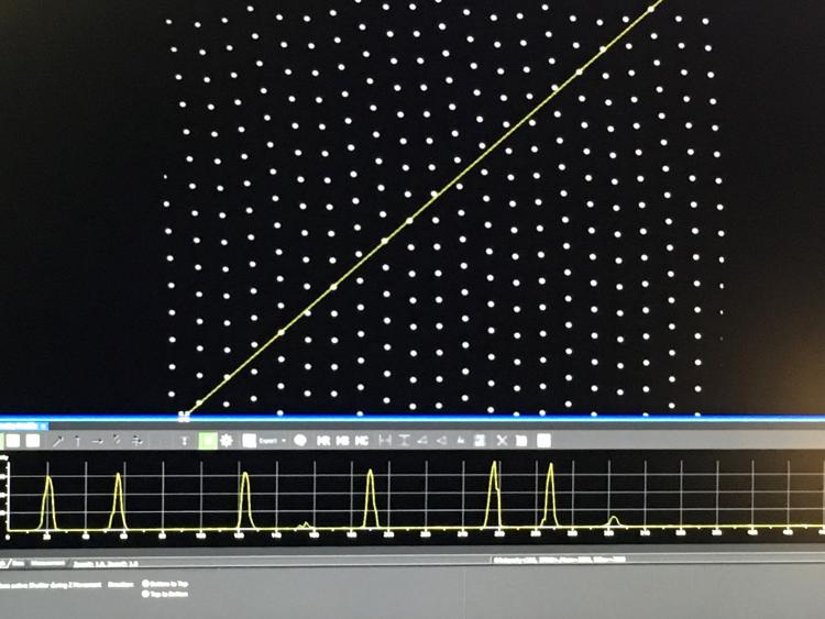
white light shining through the pinhole array (disk off)
Standard objectives
- 10x 0.45NA Plan Apo λ WD 4 (mm)
- 20x 0.75NA Plan Apo VC λ WD 1 (mm)
- 40x 0.95NA Plan Apo λ WD 0.25-0.17 (mm)
- 60x 1.40NA Plan Apo VC λ OIL WD 0.13 (mm)
- 100x 1.45NA Plan Apo λ OIL WD 0.13 (mm)
Available upon request
- 4x 0.13NA Plan Fluor WD 17.2 (mm)
- 60x 1.27NA water SR Plan Apo WD 0.17 (mm)
- Inquire with James.Orth@Colorado.edu
- Andor iXon Ultra 897 EM-CCD, 16bit, 512x512 16x16 micron pixels, binning and gain
- Hamamatsu ImagEM EM-CCD, 16bit, 512x512 16x16 micron pixels (widefield), binning and gain
Sample holders
- Slide, 35 (mm) dish, 50/60 (mm) dish
- Multiwell plate
Capabilities
- Fast Acquisition
- Multipoint, montaging, stitching
- Fast Z-sectioning via MCL Piezo Z-stage with 200 micron range
- Single point FRAP/PA with 405 nm Coherent OBIS laser (50mW)
- SOLA Light engine
- Motorized X, Y, Z
- Nikon Perfect Focus System
- TOKAI-HIT Cell Incubator; 1 X 35 (mm) dish or chamber slide (coverslip bottom).
Full system
For training email, James.Orth@Colorado.edu
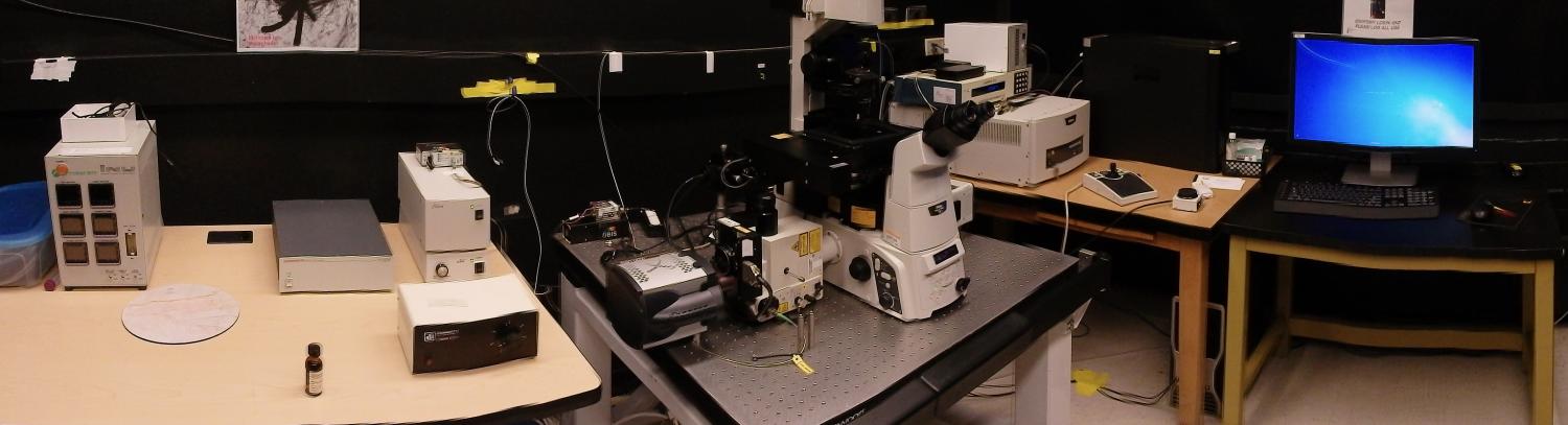
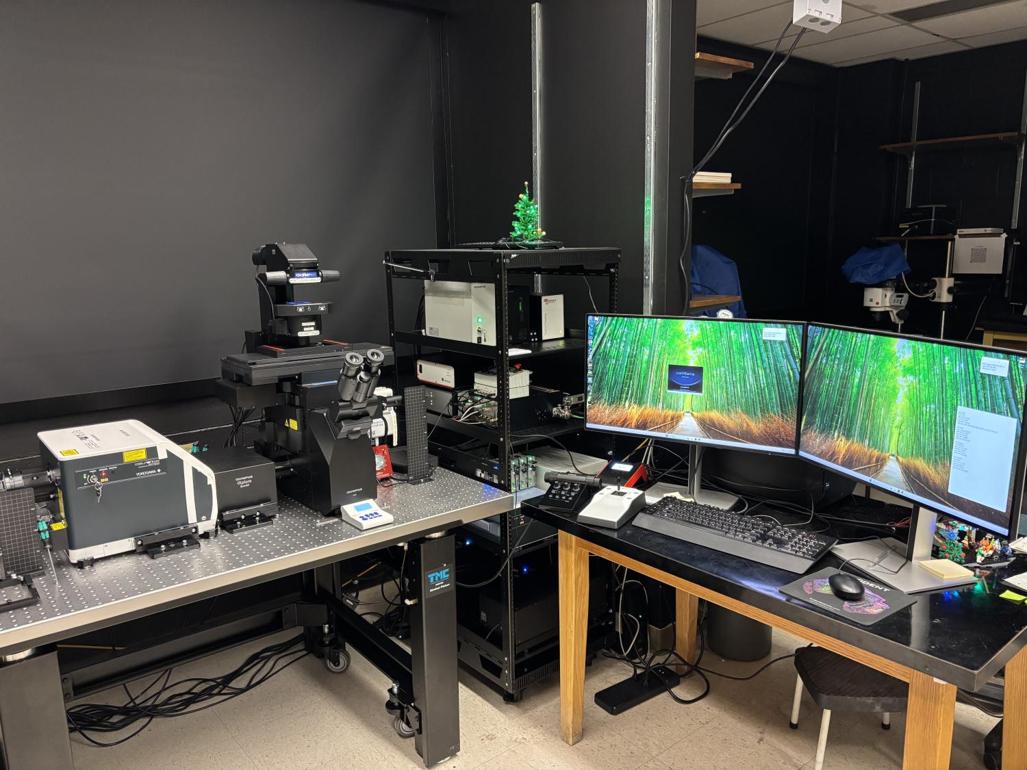
MAXIMUS - Evident Scientific IXPlore IX85 SpinSR spinning disk with super-resolution, 2 cameras and FRAP/PA, NIHS10OD038323 (Porter B059)
Lasers
- 405 (50mW), 445 (75mw), 488 (100mW), 514 (40mw), 561 (100mW) & 640 (100mW) (nm) from OBIS
Emission filters & molecules imaged (All filters are Semrock)
- Emission filter: 417-477 (nm); DAPI, Hoechst, BFP, etc..
- Emission filter: 464.5-499.5 (nm); CFP, etc.
- Emission filter: 500-550 (nm); Alexa488, FITC, EGFP, etc.
- Emission filter: 525.5-630.5 (nm); Venus, YFP, etc.
- Emission filter: 580.5-653.5 (nm); Alexa568, 594, TxRed, TRITC, mRFP, mCherry, mKO, TdTomato. etc.
- Emission filter: 665-705 (nm); Alexa647, Cy5. etc.
- Brightfield
- DIC (20, 30, 40, 60, 100x)
Dichroic spillters providing simultaneous 2 camera imaging (Semrock)
- Yokogawa CSUW1 BR488/568
- Yokogawa CSUW1P-DD03 445/514/640
Light sources and filters and viewing by eye
- CoolLED pE300Ultra; 365, 450, 560 (nm) for DAPI, GFP, RFP with triple-band emitter (Semrock)
- Lambda TLED for brightfield and DIC
- DIC (20, 30, 40, 60 and 100x)
Disks
• Yokogawa CSU-W1 Dual SoRa and 50 micron pinholes disks; W1 for spinning disk imaging, W1 and SoRa for super-resolution imaging
Standard objectives (All are Olympus)
- 4x 0.16NA UPLXAPO WD 13 (mm)
- 10x 0.40NA UPLXAPO WD 3.1 (mm)
- 20x 0.80NA UPLXAPO WD 0.6 (mm)
- 30x 1.05NA UPLSAPO SIL with correction collar, WD 0.8 (mm)
- 40x 0.95NA UPLXAPO with correction collar, WD 0.18 (mm)
- 60x 1.50NA UPLAPO OHR OIL with correction collar, WD 0.11 (mm)
- 100x 1.50NA UPLAPO OHR OIL with correction collar, WD 0.12 (mm)
Objectives available upon request
- 20x 0.45NA LUCPLFLN, with correction collar, LWD 6.4-7.6 (mm)
- 40x 0.60NA LUCPLFLN, with correction collar, LWD 2.7-4.0 (mm)
- 40x 1.30 UPLSAPO OIL, WD 0.2 (mm)
- 60x 1.20NA UPLSAPO water immersion, with correction collar, WD 0.28 (mm)
- 60x 1.35NA UPLSAPO OIL WD 0.15 (mm)
- 60x NA1.10; LUMFLN60XW LWD water immersion, with correction collar, WD 1.5 (mm)
2 ORCA-Fusion BT Gen-III sCMOS CAMERAS
- These are 2304 x 2304, 16 bit detector with 6.5 x 6.5 micron pixels
- Back-illuminated sCMOS with >90% QE from 500-650 (nm)
- Ultra-low noise
- High-speed imaging
NIH 1S10OD038323. Equipped with 2 ultra-low noise, high efficiency, Hamamatsu ORCA-Fusion BT Gen-III sCMOS cameras allows for simultaneous 2 wavelength imaging. Uses a Yokogawa W1 with 50 micron pinholes and SoRa disk for optical super-resolution. Continuous focus using Zero Drift Compensation (Olympus) and software focusing. This microscope has minimal phototoxicity and photobleaching, and is therefore great for live cell imaging at high temporal resolution, multi-point, and multi-day time-lapse. Multi-point montaging of large tissue samples in X-Y-Z.Encoded Marzhauser stage and Piezo Z stage with 500 micron range. Full DIC optics for 20, 30, 40, 60 and 100x, excellent brightfield imaging through the disk. 1X, 1.6 and 2X system magnifiers. Stage navigation and mapping. Advanced multi-point FRAP/PA using OXIS system with 100mW OBIS 405nm laser. Sample incubation and humidified atmosphere. More.
Standard sample holders
- Standard slides, multi-well slides
- 35 and 50/60 (mm) dish
- 3 slides (coming soon, custom-made)
- Multi-well plates; e.g. 6, 12, 24, 48, 96 and 384 wells
Environmentally controlled chamber
TOKAI-HIT STX with high capacity, warmed external bottle
- Single 35 (mm) dish
- Single 50/60 (mm) dish
- Chamber slide
- Multi-well plates
Acquisition
- Evident Scientific cellSens 4.4.1
Viewer
- Evident Viewer available here: Get the OlyVIA V4.2 (Build 31689) Viewer
- FIJI
Reserve the IXPLore IX85 SpinSR SDC
For training email, James.Orth@colorado.edu
CHRONOS - Yokogawa CellVoyager 1000 spinning disk confocal scanner (Porter B047A)
Lasers
- 405, 488, 561
Emission filters & molecules imaged (All filters are Semrock)
- Emission filter: 417-477 (nm) (custom); DAPI, Hoechst, BFP, etc.
- CFP has been imaged using this microscope
- Emission filter: 500-550 (nm); Alexa488, FITC, EGFP, etc.
- Venus and YFP have been imaged using this microscope
- Emission filter: 580.5-653.5 (nm); Alexa568, 594, TxRed, TRITC, mRFP, mCherry, mKO, TdTomato. etc.
- Brightfield is provided by a deep-red LED
Dichroic Mirror
- Triple, 405/388/561 (Semrock, custom)
Disks
• Yokogawa CSU-X1 Dual Nipkow disks, 25 and 50 micron pinholes - special from Yokogawa for the CV1000
Standard objectives (All are Olympus)
- 10x 0.40NA UPLAPO WD 3.1 (mm)
- 20x 0.75NA UPLSAPO WD 0.65 (mm)
- 20x 0.45NA LUCPLFLN, with correction collar, LWD 6.4-7.6 (mm)
- 40x 0.60NA LUCPLFLN, with correction collar, LWD 2.7-4.0 (mm)
- 40x 1.30NA UPLFLN OIL WD 0.2 (mm)
- 100x 1.40 UPLSAPO OIL WD 0.13 (mm)
Objectives available upon request
- 60x 1.20NA UPLSAPO water immersion, with correction collar, WD 0.28 (mm)
- 60x 1.35NA UPLSAPO OIL WD 0.15 (mm)
- 60x NA1.10; LUMFLN60XW LWD water immersion, with correction collar, WD 1.5 (mm)
- Inquire with James.Orth@Colorado.edu
Hamamatsu IMAagEM X2 1k back-thinned EMCCD high-resolution mode camera
- 1024 x 1024, 16 bit detector with 13 x 13 micron pixels
- High QE from 500-650 (nm)
- Binnig and gain to boot signal
- High-speed imaging
Chronos is equipped with an ultra-sensitive, high-resolution mode Hamamatsu EMCCD camera and a special Yokogawa X1 spinning disk with the option of dual microlens-enhanced Nipkow scanning disks with pinholes of 50 μm or 25 μm; disks can also be removed for widefield imaging. Zero Drift (Olympus) autofocusing and software focusing. This microscope has minimal phototoxicity and photobleaching, and is therefore great for live cell imaging at high temporal resolution, multi-point, and multi-day time-lapse. Sample mapping, multi-point montaging of large tissue samples in X-Y-Z and low-to-medium throughput screening. Excellent brightfield imaging through the disk. Can reload previous sample map and imaging positions to re-image the same locations at different magnifications or over time.
Standard sample holders
- Standard slides, multi-well slides
- 1 x 35 (mm) dish, incubation chamber
- 3 x 35 (mm) dish, incubation chamber
- Multi-well plates; e.g. 6, 24, 48, 96 and 384 wells
- Multi-well plates, incubation chamber; e.g. 6, 24, 48, 96 and 384 wells - must be low profile plates, contact Director for help
Environmentally controlled chambers
- Selectable temperature
- Selectable carbon dioxide concentration
- Humidified
Acquisition
- Yokogawa CV1000 1.6
Image Viewing
- Yokogawa CV1000 1.6 Viewer (PC only), contact Director
- FIJI
- Other tif viewing applications, e.g. Imaris, Nikon NIS Elements, Evident Scientific cellSens

Reserve the Yokogawa CV1000 SDC
For training email, James.Orth@Colorado.edu