
Fast-Fluorescence Microscopy Using Wavecoding Optics
Preliminary Experimental Results:
Hela cells in culture: a-tubulin labeled with FITC. (Specimen courtesy of Teresa Dibbayewan and Eleanor Kable, University of Sydney.)
Zeiss Axioplan with 40x/1.3 oil objective and cooled 12-bit Photometrics CCD (8 yrs old!).
Comparison between standard fluorescence images at 2 different planes of focus and digitally-filtered EDF images acquired from the same 2 focus planes after a CPP was installed in the microscope. Note the new EDF images are in focus over a much greater range (i.e. over 20µm as opposed to 0.5µm for the standard fluorescence images). However, some increase in noise is apparent in these "preliminary" but promising EDF results.
A confocal dataset is also shown for comparison. A focus series of 24 images, at 0.5µm steps, was averaged to form the equivalent of an EDF image. (Objective = 60x/1.4 oil). Note the similarity of axial and lateral resolution between the confocal and our new EDF images. However, this confocal image took over 20 times longer to acquire than our EDF image!
We predict we can easily increase our EDF image acquisition speed by another factor of 10 by using a more sensitive CCD camera.
Below are a few images demonstrating our results.
New EDF Fluorescence vs Standard Fluorescence of Hela Cell: FITC-tubulin.
| Standard Fluorescence | New EDF Fluroescence |
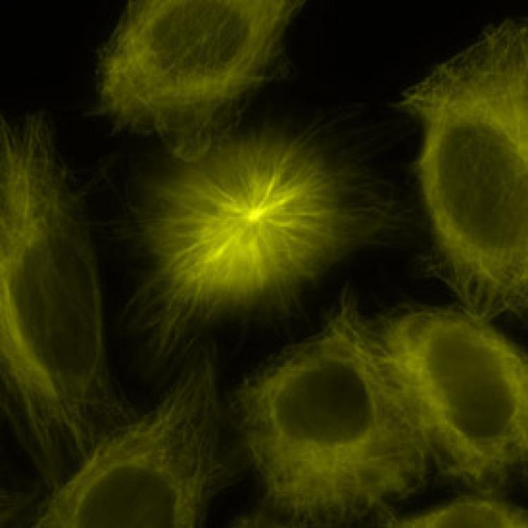 (a) One centriole of mitotic spindleis in focus, the other is blurred. Scale bar: ---- = 6µm | 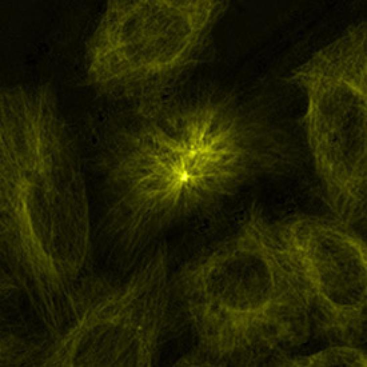 (b) EDF image shows both centrioles in focus in the 20µm |
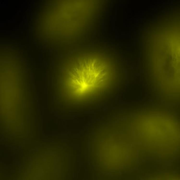 (c) Focus at 6µm below the focal plane of image (a) shows extensive blur | 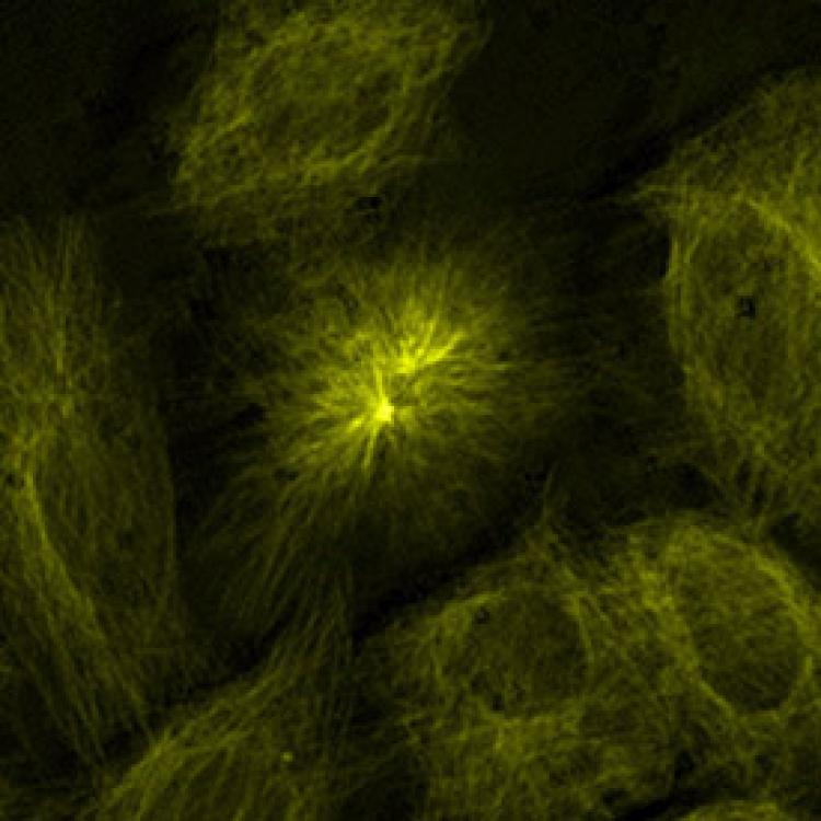 (d) Focus at 6µm below the focal plane of image(b) |
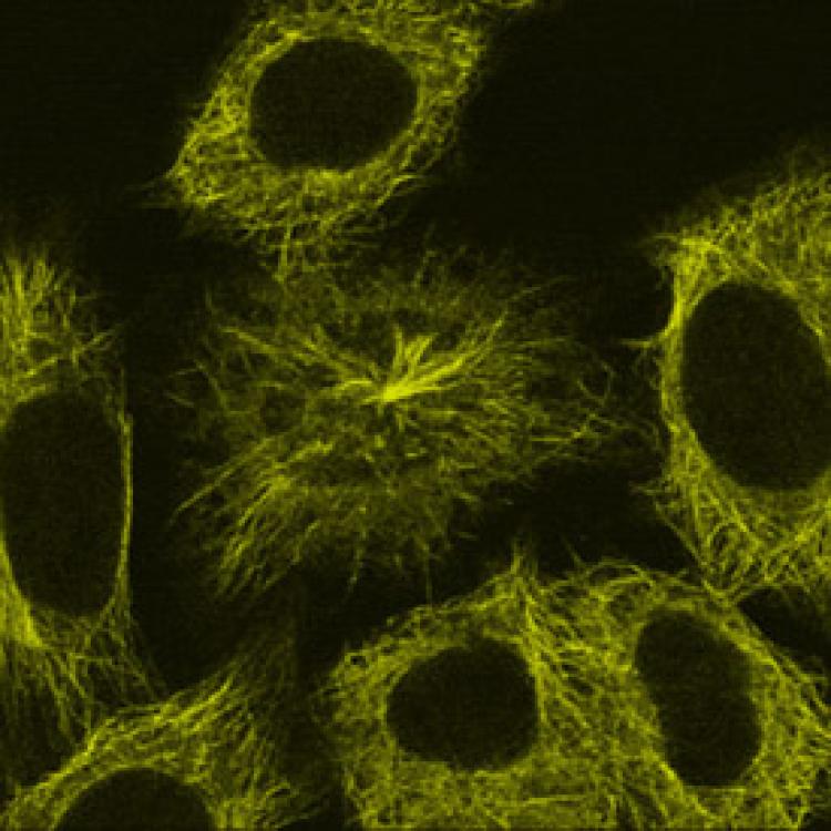
New EDF Fluorescence vs Confocal EDF Projection from 24 Planes of Focus
 exposure time = 8 seconds | 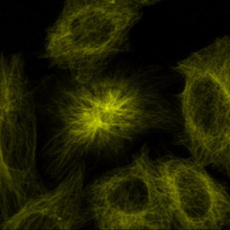 exposure time = 192 seconds |
EDF image is very similar to confocal projection image from 24 planes of focus, but EDF image was acquired in 1/20th the amount of time!! This acquisition speed will improve by at least another 10 times if a new sensitive CCD is used and the microscope illumination and optics are optimized.
Comparison of 2 different focal planes out of the 24 used in the Confocal EDF Projection
| 7th plane of focus | 15th plane of focus |
 exposure time = 8 seconds | 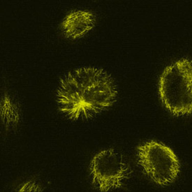 exposure time = 8 seconds |
Note our new EDF image (using an old CCD camera) was obtained in the same amount of time as one single plane from the confocal dataset!
Future Prospects:
Major advantages of our 3D high-speed fluorescence microscope:
- EDF (2D) images in milli-seconds, and 3D stereo images almost as rapidly, opening the way for studies of a wide range of live-cell or in vivo dynamics.
- Because all planes in a biological specimen volume will be illuminated and detected all at once, it should greatly reduce photo-bleaching compared to confocal and widefield systems.
- EDF microscope will require only a few modifications to existing standard commercial fluorescence microscopes - ($$$ saved!!)
E-mail questions to: carol.cogswell@colorado.edu