
Filmmakers Collaborate to Reveal New Perspectives on the Microscopic World of Bacteria
A series of films, canvases, and a dynamic living wall expose the multifaceted worlds of cyanobacteria in an exhibition now on display at the Denver Museum of Nature & Science (DMNS). REFRESH reveals microscopic landscapes that allow us to ponder how these prehistoric organisms shaped our world, and how they could help us move toward a cleaner future.
This collaboration at the intersection of biochemistry and the arts, created by faculty at the University of Colorado Boulder, provides an extraordinary view of the shape, structure, and interaction of cyanobacteria, diving into the diversity and scope of form, function, and scientific potential of these essential and complex organisms. Viewers are invited to explore the fascinating form and color of four different species of cyanobacteria across the living wall exhibit and immerse themselves in the microscopic landscapes portrayed in the films. The exhibit is free with admission, and REFRESH is located within Expedition Health on Level 2. The DMNS is open every day (9 a.m. – 5 p.m. and most Fridays until 9 p.m.), and tickets are available at www.dmns.org.
“I would love for people to marvel at these organisms, to think about the fact that we have not always had this much oxygen on the planet,” said co-creator Erin Espelie. “Cyanobacteria actively changed our atmosphere, allowing for the evolution of life. Now we are actively changing our atmosphere, and cyanobacteria could be a mechanism to counter some of the effects.”
The exhibition showcases ways in which the public can spend some time living amongst the cyanobacteria. It was sparked by the meeting of filmmakers Erin Espelie, Associate Professor in Arts & Sciences and the College of Media, Information and Communication, and Co-Director of the Nature, Environment, Science & Technology (NEST) Studio for the Arts, and Jeff Cameron, Assistant Professor in the Department of Biochemistry and a Fellow of the Renewable and Sustainable Energy Institute (RASEI; a joint institute between CU Boulder and the National Renewable Energy Laboratory (NREL)).
“The images and cinematography generated in this project are incredible, and it has been a privilege to inhabit this microscopic world,” said Espelie.
“I would love for people to marvel at these organisms, to think about the fact that we have not always had this much oxygen on the planet”
Cyanobacteria are responsible for one of the most monumental global changes in Earth’s history. Around 2.5 billion years ago The Great Oxidation Event (GOE) transitioned the earth’s atmosphere from containing almost no oxygen to containing significant levels of oxygen. This proved toxic to many anaerobic organisms which do not require molecular oxygen for growth, providing a driving force for the evolution of aerobic organisms like ourselves that do require oxygen. Evidence indicates that this transition was caused by cyanobacteria, which harnessed energy from the sun through photosynthesis to fix carbon dioxide and produce oxygen as a byproduct. The storage of chemical energy and production of oxygen led to the subsequent development of multi-cellular life-forms, forever changing the course of life on this planet.
Research suggests that cyanobacteria could be key in our efforts to regulate the levels of carbon dioxide in the earth’s atmosphere today, a challenge central to the climate crisis.
The extraordinary function of cyanobacteria is such that their impact may not just be felt in our past, but also our future. Researchers believe that these organisms could play a central role in tackling the challenges of climate change. The Cameron group use advanced microscopic imaging and analysis techniques to probe the biological machinery inside the bacteria. Of particular interest is how cyanobacteria can play a role in capturing carbon dioxide, a potent greenhouse gas and atmospheric pollutant accelerating climate change, from the air, and concentrating it to produce useful chemical products.
“Cyanobacteria have a highly efficient CO2-concentrating mechanism (CCM) that enables biological capture and conversion of carbon dioxide into useful biomolecules that can benefit society and the environment” says Cameron.
The creative minds behind this collaboration first met at a leadership workshop hosted by the CU Boulder Research & Innovation Office (RIO).
“We quickly found that we had common ground in our work; we are both filmmakers,” said Espelie.
However, the subjects of the two teams’ work are quite different; Erin’s documentary work focuses on innovative ways to ethically chronicle wildlife and the natural world, while Cameron’s work has centered around using advanced microscopy and synthetic biology to explore the world of cyanobacteria, and how these ancient organisms can be used to solve modern problems. Inspired by this intersection of interests and motivated to explore potential synergies, they successfully applied for a RIO SEED grant, which offered support and creative freedom for the teams to work together and explore cyanobacteria from a visual perspective.
“The SEED grant provided the opportunity for experimentation in an arts realm, enabling an exploration of our curiosities- something that is at the heart of both the arts and the sciences,” said Espelie.
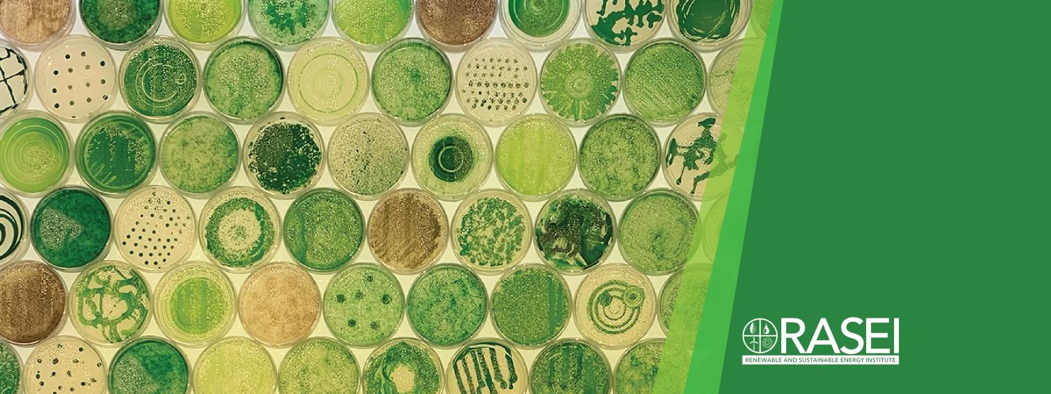
The exhibit, which opened its doors at the Denver Museum of Nature & Science in April of 2022, showcases some of this exploratory work. It features short films, still images on canvas, and artfully-designed Petri dishes of live cyanobacteria that document some of the studies made possible by this partnership.
“Cyanobacteria have a highly efficient CO2-concentrating mechanism (CCM) that enables biological capture and conversion of carbon dioxide into useful biomolecules that can benefit society and the environment”
“Filming cyanobacteria has some really unique challenges,” said Evan Johnson, a Research Assistant and Lab Manager in the Cameron group at CU Boulder.
Taking timelapse images of bacteria growing has been done before, but what is special about cyanobacteria is that they are photosynthetic, so you are using their energy source- light- to film them. The delicate biochemical machinery that captures energy from light can be damaged if too high an intensity is used. A balance must be struck: enough light to grow and visualize the bacteria, but not so much that it damages the organisms.
Scientific research can have the reputation of being rather dull and dry, full of lists of data and complex equations. This collaboration provides a window to the more exciting and colorful aspects of working in a research lab. “So often when we are looking through the microscope, we see something beautiful, but because we have a hypothesis to test or data to collect, we don’t spend time capturing these aesthetically pleasing images, and instead we focus on specific regions. This project provided opportunities to visualize the amazing microscopic world without those constraints,” said Johnson.
As a research scientist, it is these observations that spark curiosity and motivate further investigation. Often, this excitement is not captured in scientific articles and remains the exclusive domain of those working in the lab. This project bridges that gap and provides a fantastic opportunity for a wider audience to gain a glimpse of the beauty and wonder of performing scientific research.
When the teams first embarked on this project, they expected to be able to wrap things up within a year, but the synergy of the work has changed that, prompting both teams to grow and expand the scope of this project. Generating the films and images challenged the Cameron group to develop their protocols to capture longer time-lapse films, while Erin’s team underestimated the possibilities in exploring this microscopic world.
“The potential for the footage is limitless, and it is constantly changing,” said Espelie.