
Materials Research X-Ray Diffraction (XRD) Facility

The Materials Research X-Ray Diffraction (XRD) Facility at the University of Colorado Boulder provides cutting-edge capabilities for probing the structures of materials such as polymers, biological macromolecules, meso- and nano-porous materials, and molecular self-assemblies with length scales in the range 2 Å – 1000 Å using conventional and grazing-incidence SAXS and WAXS.

This Forvis instrument is built on a 20' optical table with a sliding-door radiation enclosure. The X-ray generator is a microfocus, sealed-tube source and the incident beam is collimated using multilayer mirrors and motorized, scatter-free slits. The temperature-controlled sample stage allows manual or motorized control of sample rotation, translation (x-y-z) and tilt.
Background scattering is minimized through the use of an evacuated flight path and vacuum-compatible motorized slits. Techniques that can be applied to study materials include SAXS, GISAXS, WAXS, and GIWAXS. The 30 W Xenocs Genix 3D X-ray source and state-of-the-art Dectris Eiger R 1M detector provide an excellent way to probe almost any material of interest. In-situ X-ray studies may be carried out using temperature controlled sample holders in both SAXS and GISAXS modes.
Features
- Techniques: SAXS, GISAXS, WAXS, GIWAXS
- X-ray source: 30 W Genix 3D X-ray generator (Cu anode, wavelength = 1.54 Å)
- Multilayer monochromator
- Beam size at sample: 0.8 x 0.8 mm2 for SAXS (adjustable to 0.2 x 0.8 mm2 for GISAXS)
- Optics: four sets of motorized slits for adjusting resolution
- Flight tubes: evacuated
- Detector: Dectris Eiger R 1M
- Sample holders: transmission stage with capillary holder, thin film sample holder, flat sample holder, powder sample holder
- Special stage holder: temperature-controlled (-50 to +200oC) sample holder for reflectivity (GISAXS and GIWAXS) and transmission (SAXS and WAXS) measurements
- Flux: ~ 4x10⁷ photons/s flux achieved with 0.8 x 0.8 mm2 beam
- Typical range of length scales probed: 2 Å - 1000 Å
- Computer: Linux PC
- Software for data collection: SPEC
Facility Summary
- Location: DUANE C232
- Technology focus: X-ray diffraction
- Diffractometer features:
- X-ray source: 30W Genix 3D X-ray generator
(Cu anode, wavelength = 1.54 Å)
Multilayer monochromator, evacuated flight tubes - Sample temperature control from -50 to +200ºC
- Dectris Eiger R 1M area detector
- X-ray source: 30W Genix 3D X-ray generator
- Software: spec, ImageJ/Fiji, Igor; Linux OS
- Available by appointment for research and training for CU and outside users
For more information, please contact Lab Manager Viki Martinez: xraylab@colorado.edu.
Contact Information
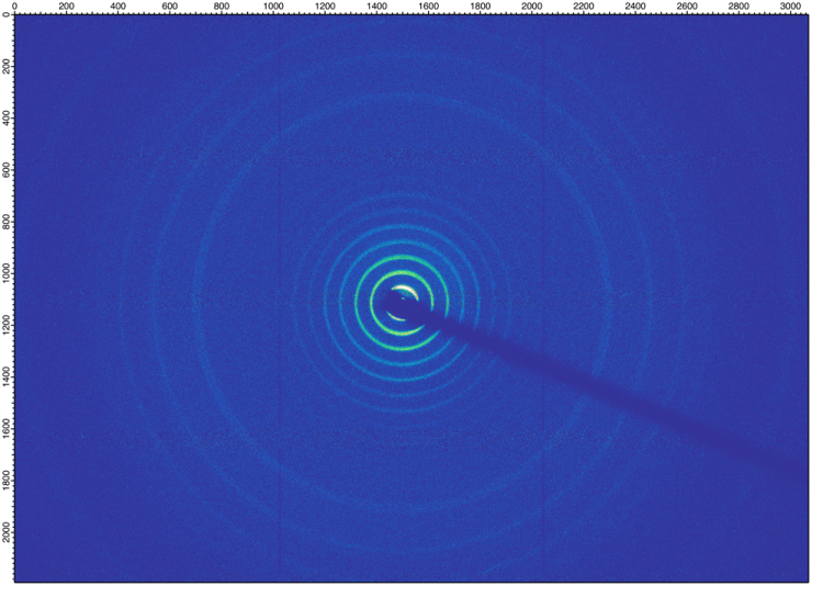
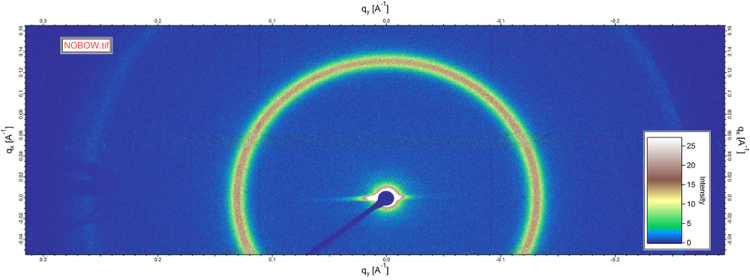
Small-angle x-ray diffraction from the bent-core liquid crystal NOBOW in the helical nanofilament phase
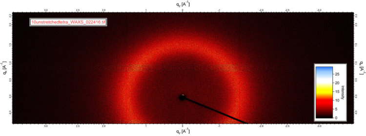
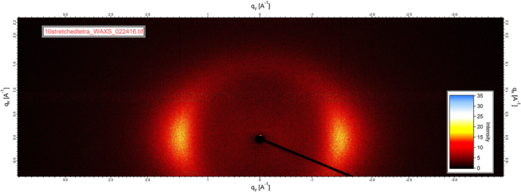
Wide-angle diffraction from a nematic liquid crystal elastomer before and after stretching
Acknowledgement
Users should include the following text in publications using data obtained in the XRD facility: "The authors gratefully acknowledge use of the Materials Research X-Ray Diffraction Facility at the University of Colorado Boulder (RRID: SCR_019304), with instrumentation supported by NSF MRSEC Grant DMR-1420736."